Panorama of Emergency Medicine

PoEM is an international peer-reviewed (double-blind) independent open access journal dedicated to advancing knowledge and practice in emergency medicine.

ISSN : 3006-0966

Skip to main navigation menu Skip to main content Skip to site footer

ED operations, triage & diagnostics

3 Items

All Items

  • Admission Criteria and Successful Care of Adults in a Tertiary Care Pediatric Hospital During the COVID-19 Pandemic

    Objective

    The study assessed the health outcomes of adult patients admitted on Pediatric Wards and Intensive Care Unit (PICU) of the Montreal Children’s Hospital (MCH) in Canada during the first and second waves of the COVID-19 pandemic.

    Methods

    This retrospective chart review analyzed data regarding all adult patients hospitalized at the MCH on pediatric units between March 14, 2020 and March 31, 2021.

    Results

    Forty (40) adults were admitted to MCH pediatric units. The median age was 28.5 years. There were 26 females and 14 males. The average length of stay (LOS) at the MCH was 6.3 days. There were 32 consultations from adult medical consultants performed at the MCH. Seventeen (17) incident reports were completed. There were no in-hospital deaths, but 6 in-hospital incidents were reported. Within the first seven days post discharge from the pediatric units, 1/24 (4%) patients returned to the adult emergency room and an additional 4 patients (16,6%) returned within 30 days.

    Conclusion

    With clear admission criteria, careful planning, and well-planned supportive resources treating adult patients within a pediatric care facility by pediatric care teams, also caring for pediatric patients in the same units, could be considered as a safe contingency plan in a time of crisis. Furthermore, admitting and treating adult patients on several pediatric units (PICU) may provide a larger variety of options for admission of adult patients.

    Introduction

         The COVID-19 pandemic put a significant stress on the healthcare systems leading to a shortage of hospital beds and equipment, disproportionally affecting older adults compared to the pediatric population1. In response to the critical rise in adult care demands, and considering that very few children required hospitalization due to COVID-19, innovative collaborations were developed to expand adult care capacity.

    Pediatric medical staff from children’s hospitals and pediatric centres worldwide stepped up to support their adult colleagues. Pediatric institutions employed different strategies to address this challenge, including the admission of adults on pediatric care units     2,3. Others adopted a hybrid model of care     4-6, or entirely repurposed their units to exclusively care for adults     7-9. In most cases, pediatric patients were either consolidated onto other units within the same hospital, transferred to standalone pediatric hospitals, or referred to community centres when specialized care was not required.

    The first COVID-19 positive case in Canada was reported on January 25, 2020, and on February 27, 2020 in Quebec10-11. On March 14 of the same year, the Quebec government declared state of health emergency12. Rapidly, Montreal became the epicentre of COVID-19 in Canada with the most cases during the first wave13. The Montreal Children’s Hospital (MCH), part of the Glen site of the McGill University Health Center (MUHC) was the first Canadian pediatric centre to admit adult patients within its own walls during the COVID-19 pandemic.

    There is limited knowledge on the occurrence of adverse events for adult patients admitted in pediatric hospitals, and on adult patients being treated by hospitalist pediatricians in Canada. 

    The objective of this study was to assess outcomes of adult patients admitted on pediatric wards and intensive care unit (PICU) of the Montreal Children’s Hospital (MCH) during the first and second waves of the COVID-19 pandemic.

    Methods

    Study Design and Setting

    In this retrospective study, a thorough medical chart review of adult patients hospitalized on the MCH units (PICU, pediatric medicine, and pediatric surgery) during the first and second wave of the COVID-19 pandemic between March 14, 2020 and March 31, 2021 was conducted. The first wave occurred between March 14, 2020 and August 31, 2021 whereas the second wave took place from September 1, 2021 through March 31, 2021. The MCH is an urban tertiary care pediatric hospital and one of the four physically joint care facilities that form the MUHC, which is situated in Montreal, Canada. Admission criteria were developed carefully during the pandemic by a multidisciplinary group of pediatric and adult health providers and were used to select the adults to be admitted on pediatric units (Table 1). 

    Data Collection

    Medical charts were reviewed by one researcher. The data was recorded in an excel spreadsheet, specifically designed for this study. The collected data included: patient demographics, primary diagnosis (as reported on the hospitalization summary), length of stay, location of the hospitalization, number of consultants from the adult medical staff involved, whether transfers occurred at any point during hospitalization on pediatric units (from pediatric wards to PICU and from pediatric units to adult units), disposition, and patient health outcomes (including: Code Blue activations, in-hospital deaths, incident reports, unplanned return visits to any of the MUHC emergency departments).

    Data analysis

    The study is descriptive, and no comparison groups were included. Descriptive statistics, encompassing the mean, standard deviation, and interquartile range (IQR), were computed to analyze various aspects of the dataset, such as patient age, length of stay and the involvement of adult consultants. Additionally, frequency tabulations were utilized to analyze primary diagnoses and patient health outcomes such as transfers, code blue activations, in-hospital deaths, incident reports, dispositions, and unplanned return visits.

    Ethical Considerations

    The MUHC Research Ethics Board approved the study protocol (#2022-7761) which was conducted in accord with the Tri-Council Policy Statement: Ethical Conduct for Research Involving Humans 2 (2018), as well as in respect of the requirements set out in the applicable standard operation procedures of the MUHC Research Institute. Anonymity and confidentiality were maintained throughout the study.

    Results

    This retrospective chart review identified a total of 40 admissions of adult patients to the MCH pediatric medical and surgical wards, as well as the PICU. These patients were cared for by a pediatric team on shared units alongside regular pediatric patients. The patients were mostly young, healthy adults, with a median age of 28.5 years. Among them, 26 were females and 14 were males (     Table 2). The average length of stay for all adult patient admissions was 6.3 days at the MCH and 22.2 days overall for those who were transferred back to the adult site when bedspace became available to complete their hospitalization.

    As demonstrated in Table 2, the 40 adult patients included in the study received a total of 132 consultations from adult medical consultants, with the number of consultations per patient ranging from 0 to 16. Of note, only 32 consultations occurred during the hospitalization on the pediatric units, divided amongst 22 patients. Among those, 15 consultations were in follow-up to the initial non-MCH phase of admission, while 17 were initiated by the pediatric medical staff to address evolving healthcare needs of patients during their stay on the pediatric units.

    Seventeen (17) incident reports were completed during admission at the MCH. There were no in-hospital deaths reported, and 6 in-hospital complications occurred: 1 line infection; 1 code blue activation; 1 accidental extubation, and 3 cases of ventilator-associated pneumonia.

    Following discharge from the MCH, most adult patients (n=24) returned home, while others were transferred to various healthcare facilities (n=16). Within 7 days post discharge from the pediatric units, 1 out of 24 patients (4%) returned to the adult emergency room, and an additional 4 patients (16.6%) returned within 30 days. Reasons for return visits varied and included new or worsening symptoms, post-operative complications, therapeutic interventions, or equipment dysfunctions. These findings are detailed in Table 4.

    Discussion

    A MUHC-wide strategy was implemented to provide the necessary capacity for adult patients while continuing to care for pediatric patients. During the first and second waves of the pandemic, this strategy involved admissions of adult patients on the PICU as well as the pediatric medicine and surgical wards of the MCH.

    The MCH opted to implement a hybrid model of care, wherein adults received treatment alongside children, administered by the same pediatric medical staff and within the same facility. Admissions originated from various sources and encompassed diverse primary diagnoses, resulting in positive and safe patient health outcomes. A pivotal factor contributing to this success was the collaborative development of clear admission criteria. Indeed, these criteria were developed by a multidisciplinary committee mandated to elaborate patient care trajectories and admission guidelines for adults on the different pediatric units (     Table 1). The selection of adult cases that were not excessively complex ensured favorable health outcomes for these adult patients admitted to pediatric units while also ensuring safety of the admitted pediatric patients.

    The study by Deep & colleagues4 described solely a hybrid pediatric and adult critical care unit without involvement of other pediatric units. The identification of adult patients for admission to the hybrid critical care unit was determined by a tactical lead from the adult team who would discuss individual patients for admission with the pediatric critical care lead. No predetermined admission criteria were reported.

    Sinha et al reported that 7 PICUs in England repurposed their space, equipment, and staff to care for a total of 145 critically ill adults during the first wave of the pandemic. The adults admitted were older (median age ranging from 57 to 62 years old in the different PICUs) as well as their in-patient mortality rate. Indeed, 20 of 145 (14%) adult patients admitted died in the PICU vs 0 in-patient deaths in all the pediatric units caring for adult patients at the Montreal Children’s hospital. In addition, the study did not publish any specific admission criteria for adults care for the 7 PICUs.

    Our experience at the MUHC reported in this study highlights both the effectiveness and safety of pediatric teams caring for adult patients in the PICU, as well as on pediatric surgical and medical wards. Collaboration and communication between the pediatric and adult medical teams as well as careful elaboration of admission criteria were instrumental in the success of the strategy.

    Whilst being very safe, the strict criteria established that adult patients infected with COVID-19 and requiring ICU level care had to be aged 30 years old or less to be considered for admission on the PICU. This significantly limited the number of potential adult PICU admissions. Return visits to the emergency room after discharge were only assessed at the MUHC and not in other nearby hospitals. In addition, the findings of this study may not apply to adult and pediatric facilities that are not located inside the same building. In such contexts, support from adult health providers may not be readily available and transport of patients from one hospital to the other may cause delays to admission.

    Conclusion

    With clear admission criteria, careful planning, and well-planned supportive resources, treating adult patients within a pediatric care facility by pediatric care teams, also caring for pediatric patients in the same units, could be considered as a safe contingency plan in a time of crisis. Furthermore, admitting and treating adult patients on several pediatric units (PICU, pediatric medicine ward, and pediatric surgical ward) may provide a larger variety of options for admission of adult patients. It would be ideal for healthcare facilities, without such experience, to prepare a similar strategy before the next pandemic strikes. Future research could assess if a similar strategy would be safe in other contexts, such as mass casualty incidents, natural, or manmade disasters.

    Acknowledgements:
    Additional contribution: We extend our appreciation to David Iannuzzi for providing research assistance during data collection, Karine Jones for her support in manuscript preparation, and Lindsay Hales for her excellent librarian support.


    Declarations

    Author contributions

    All authors contributed equally and validated the final version of record.


    Conflicts Of Interest

    The Author(s) declare(s) that there is no conflict of interest.


    Funding

    This research received no specific grant from any funding agency in the public, commercial, or not-for-profit sectors.


    Registration

    No registration applicable


    Data availability statement

    The data that support the findings of this study are available from the corresponding author upon reasonable request.


    Ethical approval

    Ethical approval for this study was not required.

    References

    1.      Guan W, Ni Z, Hu Y, Liang W, Ou C, He J, et al. Clinical Characteristics of Coronavirus Disease 2019 in China. N Engl J Med. 2020 Apr 30;382(18):1708–20. https://doi.org/10.1056/NEJMoa2002032

    2.      Dong Y, Mo X, Hu Y, Qi X, Jiang F, Jiang Z, et al. Epidemiology of COVID-19 among children in China. Pediatrics. 2020 Jun;145(6):e20200702. https://doi.org/10.1542/peds.2020-0702

    3.      Lu X, Zhang L, Du H, Zhang J, Li YY, Qu J, et al. SARS-CoV-2 Infection in Children. N Engl J Med. 2020 Apr 23;382(17):1663–5. https://doi.org/10.1056/NEJMc2005073

    4.      Deep A, Knight P, Kernie SG, D’Silva P, Sobin B, Best T, et al. A Hybrid Model of Pediatric and Adult Critical Care During the Coronavirus Disease 2019 Surge: The Experience of Two Tertiary Hospitals in London and New York. Pediatr Crit Care Med. 2021 Feb;22(2):e125–34. https://doi.org/10.1097/PCC.0000000000002584

    5.      Sinha R, Aramburo A, Deep A, Bould EJ, Buckley HL, Draper ES, et al. Caring for critically ill adults in paediatric intensive care units in England during the COVID-19 pandemic: planning, implementation and lessons for the future. Arch Dis Child. 2021 Jun 1;106(6):548–557. https://doi.org/10.1136/archdischild-2020-320962

    6.      Osborn R, Doolittle B, Loyal J. When Pediatric Hospitalists Took Care of Adults During the COVID-19 Pandemic. Hosp Pediatr. 2021 Jan 1;11(1):e15–8. https://doi.org/10.1542/hpeds.2020-001040

    7.      Yager PH, Whalen KA, Cummings BM. Repurposing a Pediatric ICU for Adults. N Engl J Med. 2020;382(22):e80. https://doi.org/10.1056/NEJMc2014819

    8.      Philips K, Uong A, Buckenmyer T, Cabana MD, Hsu D, Katyal C, et al. Rapid Implementation of an Adult Coronavirus Disease 2019 Unit in a Children’s Hospital. J Pediatr. 2020 Jul 1;222:22–7. https://doi.org/10.1016/j.jpeds.2020.04.060

    9.      Joyce CL, Howell JD, Toal M, Wasserman E, Finkelstein RA, Traube C, et al. Critical Care for Coronavirus Disease 2019: Perspectives From the PICU to the Medical ICU. Crit Care Med. 2020;48(11):1553–5. https://doi.org/10.1097/CCM.0000000000004543

    10.  Berry I, Soucy JPR, Tuite A, Fisman D. Open access epidemiologic data and an interactive dashboard to monitor the COVID-19 outbreak in Canada. CMAJ. 2020 Apr 14;192(15):E420. https://doi.org/10.1503/cmaj.75262

    11.  Santé et Services sociaux. Cas confirmé de COVID-19 au Québec [Internet]. Quebec: Gouvernement du Québec; 2020 [cited 2021 Mar 1]. Available from: https://www.quebec.ca/nouvelles/actualites/details/cas-confirme-de-covid-19-au-quebec

    12.  Santé et Services sociaux. Pandémie de COVID-19 - Le gouvernement du Québec déclare l’état d’urgence sanitaire, interdit les visites dans les centres hospitaliers et les CHSLD et prend des mesures spéciales pour offrir des services de santé à distance [Internet]. Quebec: Gouvernement du Québec; 2020 [cited 2021 Mar 1]. Available from: https://www.quebec.ca/nouvelles/actualites/details/pandemie-de-covid-19-le-gouvernement-du-quebec-declare-letat-durgence-sanitaire-interdit-les-visites-dans-les-centres-hospitaliers-et-les-chsld-et-prend-des-mesures-speciales-pour-offrir-des-services-de-sante-a-distance

    13.  Health Canada [Internet]. Quebec: Government of Canada; 2020. Epidemiological summary of COVID-19 cases in Canada. Available from: https://health-infobase.canada.ca/covid-19/epidemiological-summary-covid-19-cases.html

  • Risk Factors for Return Visits to the Pediatric Emergency Department: Systematic Search and Review

    Objectives: Return visits (RVs) to the emergency department (ED) has always been a major concern. RVs to the emergency department are a big burden on the healthcare system as its cost is higher than the cost of the initial visit. This review was performed to identify factors associated with risk of RVs to the pediatric ED.

    Methods and Analysis: One researcher searched Medline, Embase, Cochrane Library and Web of Science. Studies were identified by using MeSH and keywords and included RVs to the pediatric ED up to 1 year a primary outcome. All studies were screened by two independent reviewers for eligibility and in case of disagreement, a meeting was held to discuss the problematic studies and a consensus was achieved.

    Results: The search identified 539 reports from which 28 articles were included. Data was then extracted from the included studies according to a preset format. The exposures were grouped in 3 different groups: very probable, possible, and less likely.
    As a result, young age, language barrier and high acuity were identified as very probable risk factors. Having a public insurance or with low income, patients with comorbidities and patients who had multiple previous ED visits were found to be possible risk factors for return visits.

    Conclusion: Young age, high acuity and language barrier among others are risk factors for return visits to the pediatric ED. Physicians should be aware of these factors and have a low threshold for admission or a good discharge plan for patients with one or more factors.

    Key Messages:

    Ø  What is already known on this topic – Return visits (RV) to the pediatric emergency department has always been a major concern and a big burden on the healthcare system as its cost is higher than the cost of the initial visit. The risk factors for RV vary widely. 
    Ø  What this study adds – Young age, language barrier and high acuity were identified as very probable risk factors. Having a public insurance or with low income, patients with comorbidities and patients who had multiple previous ED visits were found to be possible risk factors for return visits.
    Ø  How this study might affect research, practice, or policy – Physicians should be aware of these factors and have a low threshold for admission or a good discharge plan for patients with one or more factors.

    Introduction
         Return visits (RVs) to the emergency department (ED) have always been a major concern. In fact, since the 1980s, emergency physicians recognized return visits, also known as bounce back visits, as a “red flag” for low quality of care [1]. In general, RVs to the emergency (ED) constitute an enormous burden on the healthcare system. For instance, on a financial level, the cost of a RV is higher than the cost of the initial visit [2]. Further, on a medical level, patients admitted to the Pediatric Intensive Care Unit (PICU) following a RV to the pediatric ED, are more likely to be put on a ventilator [3]. For these reasons, RVs have often been used as a quality metric in the pediatric ED [4]. 

         As traditionally reported in many studies, a visit occurring within 72-hours of an index presentation for the same complaint is considered to be a RV because it reflects either an inadequate treatment or a missed diagnosis [5-7]. However, more recently, the 72-hour limit has been challenged by many as it might not mirror neither of both classifications (i.e., inadequate treatment or missed diagnosis) [8]. Indeed, some of the most common pediatric presentations to the ED, such as allergic reaction, asthma, fever, and bronchiolitis, can either deteriorate or have symptoms requiring a RV to the ED after more than 3 days, which in this case, doesn’t constitute a low quality of care. Longer time periods have also been proposed in adult EDs. For example, a recently published large retrospective study including more than one million adult ED RVs over 10 years used an upper limit of 14 days after the initial visit. The authors concluded that these patients should be identified early to avoid intensive care unit (ICU) admission during the RV [9].

         Identifying risk factors leading to RVs could contribute to improved care for children presenting to pediatric EDs. An initial search performed in preparation for our study identified one literature review published in 2016 on the topic of risk factors and interventions that affected RVs to the pediatric ED [8]. This review concluded that mental health problems, younger age, acuity of illness, medical history of asthma, and social factors are risk factors for RVs. However, this review looked at studies published prior to November 2012 and identified only 6 studies looking at RVs ranging from 48 hours to 1 year. Also, as the authors mentioned in their study limitations’ section, they only investigated Medline without grading the reviewed studies for quality. Further, 3 out of the 6 studies were limited to a specific condition (2 included only patients with asthma and 1 included patients with mental health-related issues), limiting therefore the diversity of the collected data and hence its generalizability to the context of RVs. 

         To fill out the previous gap, we performed a systematic search and review to identify factors associated with risk of RVs to the pediatric ED in children of any age as defined by the authors in each article. As there is no true consensus on the delay where a subsequent visit would be considered a RV, we considered 1 year as a reasonable cutoff as considered in the previous review mentioned prior. However, no differentiation was made between RVs for the same problem and RVs for an unrelated complaint to the initial visit. We aimed to answer the following question: in pediatric patients of any age presenting to the emergency department, what risk factors on the first presentation would predict a RV within 1 year?

    Materials and Methods

    Search Strategy
    The following databases were searched for relevant records on April 22 2021: Medline (via Ovid 1946 to 2021 April 21), Cochrane (via Wiley, from the Cochrane Database of Systematic Reviews, Issue 3 of 12, March 2021), Embase (via Ovid 1947 to 2021 April 21), and Web of Science (via Clarivate, Indexes SCI-EXPANDED, SSCI, A&HCI, CPCI-S, CPCI-SSH, BKCI-S, BKCI-SSH, ESCI, CCR-EXPANDED, & IC).
    The search strategies designed by a librarian (AB) used text words and relevant indexing to identify records on risk factors for pediatric emergency readmission. 
    The final Medline strategy (Appendix 1) was adapted for all databases, with modifications to search terms and syntax as necessary. No language limits were applied. 

    Inclusion Criteria
    The included studies were limited to randomized controlled trials (RCTs), controlled studies, systematic reviews, cohort studies, case control studies and cross-sectional studies written in English or French without any date limit for inclusion. Case reports and series were excluded. Also, studies looking into one diagnosis (like asthma, bronchiolitis…) were excluded because risk factors for RVs can be confounding factors and not true risk factors (for example, bronchiolitis is diagnosed in kids less than 12-24 months making young age a confounding factor), however, studies with a large scope of presentation and low risk for confounding factors (like trauma) were included.
    No restriction was made on the past medical history, perinatal history, previous hospitalizations, medications, gender, acuity on presentation, medical interventions on the first visit, length of stay (LOS), left without being seen (LWBS), left against medical advice (AMA) or any other criteria. However, according to our search, these might be identified as risk factors for RVs.

    Screening
    All studies were screened by title first then by abstract by two independent reviewers (CET and IC). Duplicated studies in different databases were removed. Studies not satisfying the inclusion criteria were then excluded. Further, following an independent extraction of data, both reviewers compared results to reach a consensus. 

    Data extraction
    Data were extracted by the two independent reviewers according to a predefined format (Table 1). Data points collected included the first author’s name, the study characteristics (type of study, date of publication, total number of RVs and country where the study was conducted), the timing of the RV (i.e., outcome) and the identified risk factors (i.e., exposures).

    Results

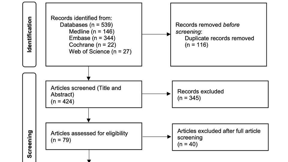
    The initial search identified 539 articles (Figure 1). 
         After removal of duplicate studies and screening of titles and abstracts by the 2 independent reviewers, 79 articles were included. Forty reports were then excluded after full manuscript review by both reviewers. For the remaining articles, a meeting between the 2 reviewers and a consensus to include 28 articles was achieved [8, 10-36].

         Data were then extracted from the included studies according to the preset format (Table 1). The data in italic format in the table represent a negative or neutral finding (for example, in Daymont et al. discharge heart rate was not found to affect RVs). The risk factors were then divided into 3 different groups according to how many times they were cited as there was no other practical way to weigh and compare the studies (Figure 2). 


    Discussion

    This review identified multiple risk factors for RVs to the pediatric ED. 

    Young Age
    Infant and young children were, by far, more likely to have a RV to the ED than older children and adolescents even though the definition of young age was different between studies ranging from younger than 1 year to younger than 5 years. While these RVs might be due to progression of the disease, such as bronchiolitis, in this age group, clear discharge instructions and explanation of  signs of deterioration were found to be a protective factor against unnecessary RVs [37].

    Language
    Language barrier between patients and healthcare providers has been associated with increased pediatric ED visit length of stay and resource utilization in addition to increased RVs [38, 39 ]. With the increased number of refugees worldwide, language barrier is becoming more relevant and the need for multilingual healthcare professionals is constantly increasing [40].

    High Acuity
    Pediatric patients who are sicker at triage are more likely to be admitted especially in a crowded ED [41]. If those patients with higher acuity are discharged from the ED, they are more likely to bounce back. Physicians should probably maintain a low threshold for admission at the time of the initial visit [42].

    Other Risk Factors 
    This review identified more risk factors like patients with public insurance or with low income, patients with comorbidities and patients who had multiple previous ED visits. How to classify a RV after LWBS and, to a lesser extent AMA, is controversial. It might be considered as the first visit and not a RV since the patient was not assessed by the physician and/or did not receive the proper management for their condition.

    Studies Quality and Limitations
         Most of the studies were retrospective (86%) with only two prospective studies, one of which was a planned secondary analysis of a prospective cohort. The majority were North American studies (68%), limiting therefore the diversity and generalizability of our findings. The primary outcome was different across the articles: although it was mostly risk factors for RVs, there was a big discrepancy in the risk factors studied as some of the risk factors were frequent, cited up to 16 times (age), while others were rare, with only 2 citations (season).
         Due to these limitations and the heterogeneity of the articles, the wide variety of variables, as well as a lack of a true definition of the outcome (RV ranged from 48 hours to 1 year), and of a grading system for most observational studies in this review, a systematic review and meta-analysis were not feasible. Therefore, a systematic search and review was conducted [43]. Most of the articles were also retrospective and arguably have low weight to form a robust meta-analysis. In addition, RV can have different expressions ranging from unplanned revisits to readmissions visits, among others. This means that despite the comprehensive search (Appendix 1), we might have missed some articles. However, this would probably not affect the results significantly as there is a visible consensus between the different studies that were included. Also, using such a wide time limit of one year and not differentiating between RVs for the same problem and RVs for an unrelated complaint to the initial visit might have influenced the results however, most studies did not specify if the RV complaint was different from the initial visit. Finally, this review did not look at the disposition of patients during the RV as the RV outcome can range from discharging the patient home to admission to wards/ICU to mortality and would obviously affect the weight and importance of each identified risk factor. 

    Conclusion
        Young age, high acuity at presentation and language barrier, among others, are risk factors for return visits to the pediatric ED. Physicians should be aware of these factors and have a low threshold for admission or a good discharge plan for patients with one or more factors to improve quality care in the pediatric ED. While pediatric ED overcrowding is a burden worldwide, such studies can help decompress the EDs by identifying high risk patients and reducing RVs.

    Declarations
    Author contributions: All authors contributed equally and validated the final version of record.

    Conflicts Of Interest: The Author(s) declare(s) that there is no conflict of interest.

    Funding: This research received no specific grant from any funding agency in the public, commercial, or not-for-profit sectors.

    Registration: No registration applicable

    Data availability statement: The data that support the findings of this study are available from the corresponding author upon reasonable request.

    Ethical approval: Ethical approval for this study was not required.

    References

    1.      Lerman B, Kobernick MS. Return visits to the emergency department. J Emerg Med. 1987 Sep 1;5(5):359–62. https://doi.org/10.1016/0736-4679(87)90138-7.

    2.      Duseja R, Bardach NS, Lin GA, Yazdany J, Dean ML, Clay TH, et al. Revisit Rates and Associated Costs After an Emergency Department Encounter. Ann Intern Med. 2015 Jun 2;162(11):750–6. https://doi.org/10.7326/M14-1616.

    3.      Chiang CY, Cheng FJ, Huang YS, Chen YL, Wu KH, Chiu IM. ICU admission following an unscheduled return visit to the pediatric emergency department within 72 hours. BMC Pediatrics. 2019 Aug 2;19(1):268. https://doi.org/10.1186/s12887-019-1644-y.

    4.      Ostrow O, Zelinka A, Shim A, Azmat SK, Masood S, Chartier LB. Pediatric Emergency Department Return Visits: An Innovative and Systematic Approach to Promote Quality Improvement and Patient Safety. Pediatr Emerg Care. 2020;36(12):e726–31. https://doi.org/10.1097/PEC.0000000000001999.

    5.      Ko M, Lee Y, Chen C, Chou P, Chu D. Incidence of and Predictors for Early Return Visits to the Emergency Department: A Population-Based Survey. Medicine. 2015;94(43):e1770. https://doi.org/10.1097/MD.0000000000001770.

    6.      Benbassat J, Taragin M. Hospital Readmissions as a Measure of Quality of Health Care: Advantages and Limitations. Arch Intern Med. 2000 Apr 24;160(8):1074–81. https://doi.org/10.1001/archinte.160.8.1074.

    7.      Trivedy CR, Cooke MW. Unscheduled return visits (URV) in adults to the emergency department (ED): a rapid evidence assessment policy review. Emerg Med J. 2015 Apr 1;32(4):324. https://doi.org/10.1136/emermed-2013-202719.

    8.      Tran QK, Bayram JD, Boonyasai RT, Case MA, Connor C, Doggett D, et al. Pediatric Emergency Department Return: A Literature Review of Risk Factors and Interventions. Pediatr Emerg Care. 2016;32(8):570–7. https://doi.org/10.1097/PEC.0000000000000876.

    9.      Aaronson E, Jansson P, Wittbold K, Flavin S, Borczuk P. Unscheduled return visits to the emergency department with ICU admission: A trigger tool for diagnostic error. Am J Emerg Med. 2020 Aug 1;38(8):1584–7. https://doi.org/10.1016/j.ajem.2019.158430.

    10.  Ehwerhemuepha L, Yu PT, Guner YS, Wallace E, Feaster W. A Nested Mixed Effects Multicenter Model Examining the Risk Factors for Pediatric Trauma Return Visits Within 72 Hours. J Surg Res. 2021 Jan 1;257:370–8. https://doi.org/10.1016/j.jss.2020.08.021.

    11.  Pershad J, Jones T, Harrell C, Ajayi S, Giles K, Cross C, et al. Factors Associated With Return Visits at 7 Days After Hospital Discharge. Hosp Pediatr. 2020 Apr 1;10(4):353–8. https://doi.org/10.1542/hpeds.2019-0207.

    12.  Türe E, Yazar A. Retrospective Evaluation of Return Visits to the Paediatric Emergency Department. Eurasian J Emerg Med. 2020 Jun 22;19(2):115–20. https://doi.org/10.4274/eajem.galenos.2019.68926.

    13.  Drouin O, D’Angelo A, Gravel J. Impact of wait time during a first pediatric emergency room visit on likelihood of revisit in the next year. Am J Emerg Med. 2020 May 1;38(5):890–4. https://doi.org/10.1016/j.ajem.2019.07.005.

    14.  Daymont C, Balamuth F, Scott HF, Bonafide CP, Brady PW, Depinet H, et al. Elevated Heart Rate and Risk of Revisit With Admission in Pediatric Emergency Patients. Pediatr Emerg Care. 2021 Apr;37(4):e185–91. https://doi.org/10.1097/PEC.0000000000001552.

    15.  de Vos-Kerkhof E, Geurts DHF, Steyerberg EW, Lakhanpaul M, Moll HA, Oostenbrink R. Characteristics of revisits of children at risk for serious infections in pediatric emergency care. Eur J Pediatr. 2018 Apr 1;177(4):617–24. https://doi.org/10.1007/s00431-018-3095-0.

    16.  Kim BS, Kim JY, Choi SH, Yoon YH. Understanding the characteristics of recurrent visits to the emergency department by paediatric patients: a retrospective observational study conducted at three tertiary hospitals in Korea. BMJ Open. 2018 Feb 1;8(2):e018208. https://doi.org/10.1136/bmjopen-2017-018208.

    17.  Michelson KA, Lyons TW, Bachur RG, Monuteaux MC, Finkelstein JA. Timing and Location of Emergency Department Revisits. Pediatrics. 2018 May 1;141(5):e20174087. https://doi.org/10.1542/peds.2017-4087.

    18.  Meyer-Macaulay CB, Truong M, Meckler GD, Doan QH. Return visits to the pediatric emergency department: A multicentre retrospective cohort study. CJEM. 2018;20(4):578–85. https://doi.org/10.1017/cem.2017.40.

    19.  Ruttan T, Lawson KA, Piper K, Wilkinson M. Risk Factors Associated With Emergency Department Return Visits Following Trauma System Discharge. Pediatr Emerg Care. 2018;34(3):202–7. https://doi.org/10.1097/PEC.0000000000001182.

    20.  Hu YH, Tai CT, Chen SCC, Lee HW, Sung SF. Predicting return visits to the emergency department for pediatric patients: Applying supervised learning techniques to the Taiwan National Health Insurance Research Database. Comput Methods Programs Biomed. 2017 Jun 1;144:105–12. https://doi.org/10.1016/j.cmpb.2017.03.022.

    21.  Kilicaslan O, Sönmez FT, Gunes H, Temizkan RC, Kocabay K, Saritas A. Short Term Unscheduled Revisits to Paediatric Emergency Department - A Six Year Data. J Clin Diagn Res. 2017 Mar 1;11(3):SC12-SC15. https://doi.org/10.7860/JCDR/2017/25098.9484.

    22.  Samuels-Kalow ME, Stack AM, Amico K, Porter SC. Parental Language and Return Visits to the Emergency Department After Discharge. Pediatr Emerg Care. 2017 Jun;33(6):402-404. https://doi.org/10.1097/PEC.0000000000000592.

    23.  Wilson PM, Florin TA, Huang G, Fenchel M, Mittiga MR. Is Tachycardia at Discharge From the Pediatric Emergency Department a Cause for Concern? A Nonconcurrent Cohort Study. Ann Emerg Med. 2017 Sep;70(3):268-276.e2. https://doi.org/10.1016/j.annemergmed.2016.12.010.

    24.  Goh GL, Huang P, Kong MC, Chew SP, Ganapathy S. Unplanned reattendances at the paediatric emergency department within 72 hours: a one-year experience in KKH. Singapore Med J. 2016;57(6):307-13. https://doi.org/10.11622/smedj.2016105.

    25.  Schneider M, Chen C, Menoch M, Levasseur K. Primary language and return visits in the pediatric emergency department. SAEM Annual Meeting Abstracts. Acad Emerg Med. 2016 May 1;23(S1):S103-4. https://doi.org/10.1111/acem.12974.

    26.  de Vos-Kerkhof E, Geurts DH, Wiggers M, Moll HA, Oostenbrink R. Tools for 'safety netting' in common paediatric illnesses: a systematic review in emergency care. Arch Dis Child. 2016 Feb;101(2):131-9. https://doi.org/10.1136/archdischild-2014-306953.

    27.  Saunders N, To T, Parkin P, Guttmann A. The relationship between immigrant status and pediatric emergency department return visits. Paediatrics and Child Health (Canada). 2015;20:e93. https://doi.org/10.1093/pch/20.5.e93.

    28.  Sung SF, Liu KE, Chen SC, Lo CL, Lin KC, Hu YH. Predicting Factors and Risk Stratification for Return Visits to the Emergency Department Within 72 Hours in Pediatric Patients. Pediatr Emerg Care. 2015 Dec;31(12):819-24. https://doi.org/10.1097/PEC.0000000000000417.

    29.  Gallagher RA, Porter S, Monuteaux MC, Stack AM. Unscheduled return visits to the emergency department: the impact of language. Pediatr Emerg Care. 2013 May;29(5):579-83. https://doi.org/10.1097/PEC.0b013e31828e62f4

    30.  Samuels-Kalow ME, Stack AM, Amico K, Porter SC. The association between parental language and 72-hour revisits following pediatric emergency department discharge. SAEM Annual Meeting Abstracts. Acad Emerg Med. 2013 May 1;20(s1):S188. https://doi.org/10.1111/acem.12115.

    31.  Gaucher N, Bailey B, Gravel J. Impact of physicians' characteristics on the admission risk among children visiting a pediatric emergency department. Pediatr Emerg Care. 2012 Feb;28(2):120-4. https://doi.org/10.1097/PEC.0b013e318243f8e0.

    32.  Reinke DA, Walker M, Boslaugh S, Hodge D 3rd. Predictors of pediatric emergency patients discharged against medical advice. Clin Pediatr (Phila). 2009 Apr;48(3):263-70. https://doi.org/10.1177/0009922808323109.

    33.  Costabel S, Piccotti E, Sartini M, Magnani M, Di Pietro P. Return visits to the Paediatric Emergency Department: first analysis in Italy. J Prev Med Hyg. 2008 Dec;49(4):142-7. https://doi.org/10.15167/2421-4248/jpmh2008.49.4.133

    34.  Goldman RD, Ong M, Macpherson A. Unscheduled return visits to the pediatric emergency department-one-year experience. Pediatr Emerg Care. 2006 Aug;22(8):545-9. https://doi.org/10.1097/01.pec.0000230553.01917.05.

    35.  LeDuc K, Rosebrook H, Rannie M, Gao D. Pediatric emergency department recidivism: demographic characteristics and diagnostic predictors. J Emerg Nurs. 2006 Apr;32(2):131-8. https://doi.org/10.1016/j.jen.2005.11.005.

    36.  Alessandrini EA, Lavelle JM, Grenfell SM, Jacobstein CR, Shaw KN. Return visits to a pediatric emergency department. Pediatr Emerg Care. 2004 Mar;20(3):166-171. https://doi.org/10.1097/01.pec.0000117924.65522.a1.

    37.  Navanandan N, Schmidt SK, Cabrera N, Topoz I, DiStefano MC, Mistry RD. Seventy-two-hour Return Initiative: Improving Emergency Department Discharge to Decrease Returns. Pediatr Qual Saf. 2020 Sep 25;5(5):e342. https://doi.org/10.1097/pq9.0000000000000342.

    38.  Hampers LC, Cha S, Gutglass DJ, Binns HJ, Krug SE. Language barriers and resource utilization in a pediatric emergency department. Pediatrics. 1999 Jun;103(6):1253-6. https://doi.org/10.1542/peds.103.6.1253.

    39.  Zamor R, Byczkowski T, Zhang Y, Vaughn L, Mahabee-Gittens EM. Language Barriers and the Management of Bronchiolitis in a Pediatric Emergency Department. Acad Pediatr. 2020 Apr;20(3):356-363. https://doi.org/10.1016/j.acap.2020.01.006.

    40.  Hampers LC, McNulty JE. Professional interpreters and bilingual physicians in a pediatric emergency department: effect on resource utilization. Arch Pediatr Adolesc Med. 2002 Nov;156(11):1108-13. https://doi.org/10.1001/archpedi.156.11.1108.

    41.  Doan Q, Wong H, Meckler G, Johnson D, Stang A, Dixon A, et al. The impact of pediatric emergency department crowding on patient and health care system outcomes: a multicentre cohort study. CMAJ. 2019 Jun 10;191(23):E627-35. https://doi.org/10.1503/cmaj.181426.

    42.  Al-Qahtani MH, Yousef AA, Awary BH, Albuali WH, Al Ghamdi MA, AlOmar RS, et al. Characteristics of visits and predictors of admission from a paediatric emergency room in Saudi Arabia. BMC Emerg Med. 2021 Jun 21;21(1):72. https://doi.org/10.1186/s12873-021-00467-7.

    43.  Grant MJ, Booth A. A typology of reviews: an analysis of 14 review types and associated methodologies. Health Info Libr J. 2009 Jun;26(2):91-108. https://doi.org/10.1111/j.1471-1842.2009.00848.x.


  • Heart rate, a poor predictor of Pulmonary Embolism

    Objective: To determine if there is a significant difference in vital signs between patients with confirmed and excluded pulmonary embolism (PE) throughout their Emergency Department presentation.

    Methods: We conducted a retrospective cohort study with patients presenting with suspected PE to Monash Health Emergency Departments between July 2014 and July 2019. Vital signs were compared between patients with confirmed or excluded PE as determined by imaging (CTPA or VQ). Vital signs were compared at three unique data points: initial, minimum, and maximum values.

    Results: 3549 patients met inclusion criteria, 922 with confirmed PE and 2627 with excluded PE based on CTPA or VQ. Patients with PE had significant elevations in mean respiratory rates, systolic blood pressures and reduced oxygen saturations compared to patients without PE. Heart rate was not significantly different at initial and maximum datapoints.

    Conclusion: Vital signs were demonstrated to be poor predictors of acute PE. Receiver operating characteristic curve analysis suggests that heart rate has poor discriminative power. AUC values for heart rate were: 0.516 (initial), 0.549 (maximum) and 0.519 (minimum). Furthermore, 95% of patients with confirmed PE did not exceed heart rates of 100 BPM during presentation to Emergency. The utility of elevated heart rate and other vital signs in predicting PE were not substantiated in this study.


    Introduction
    A pulmonary embolism (PE) refers to a blockage in the lung’s arterial network due to the migration of clot material (1-2). Although uncommon, with a reported incidence of 60 to 70 per 100,000, mortality rates can range from up to 1% for small PEs, and between 18 to 65% in massive PEs (1). Patient with suspected PE report symptoms of dyspnoea, haemoptysis, and pleuritic chest pain. On examination, patients may also exhibit abnormal vital signs, such as tachycardia (3,4), tachypnoea (5) and hypotension (5, 6). 20 per cent of patients with suspected PE return positive diagnoses, hence, the diagnostic workflow for PE must employ safe, timely and primarily non-invasive methods (7).

    Definitive investigations for PE may include a ventilation-perfusion scintigraphy (V/Q scan) or computed tomography pulmonary angiography (CTPA) (2). To minimise inappropriate use, risk stratification tools are utilised to exclude PE in low-risk patients. These include the Wells’ criteria, revised Geneva score (rGeneva) and PERC rule, all of which employ vital signs to stratify risk of PE (8-11).

    The Wells’ criteria have been validated in numerous clinical settings to provide an estimated pre-test probability and risk stratification for PE – assisting clinicians in selecting appropriate investigations (10). To establish pre-test probability, the Wells’ criteria allocate points to clinical factors, such as tachycardia (>100 BPM) and evidence of deep vein thrombosis (DVT) (12). The rGeneva similarly quantifies risk but enables a finer level of stratification by ascribing greater weight to heart rates (HR) exceeding 95 BPM, compared to 75-94 BPM. The PERC rule increases suspicion for PE in patients with HR greater than 100 BPM and oxygen saturations less than 95 per cent (8). However, a meta-analysis demonstrated the inadequacy of these scores in the final exclusion of PE (13).

    Non-specific tachycardia in emergency department (ED) patients has reportedly led to false positive screening and unnecessary diagnostic tests (14, 15). Despite associations between abnormal vital signs and PE, these derangements are unpredictable, transient, and may even normalise during an ED stay. Thus, for a theoretically accurate stratification of risk, vital signs need to be robust and persistent, suggesting potentially limited clinical utility.

    Mortality rates in patients with confirmed PE can be estimated with the Pulmonary Embolism Severity Index (PESI) and BOVA score (16, 17). These tools utilise vital signs to inform the necessity of inpatient management. The BOVA score utilises HR and systolic blood pressure (SBP), whilst the PESI utilises HR, SBP, respiratory rate (RR), temperature, and oxygen saturation (16, 17). These scores also rely on the persistence of deranged vital signs, which suggests that they are a potentially inaccurate representation of a patient’s evolving clinical state. Hence, appraising these scores against the stability and trend of a patient’s vital signs is necessary.

    The adoption of the D-dimer was thought to revolutionise the diagnostic approach for PE and reduce unnecessary diagnostic imaging. However, as the D-dimer has inherently low specificity and excellent sensitivity, there is potential for false positivity that has been criticised in the literature (18-20). There is an evolving body of research focused on advancing and optimising the diagnostic approach for PE, resulting in novel technologies, such as focused cardiac ultrasound. However, current practice continues to place significance on vital sign derangements, which could potentially impede development of novel clinical approaches (21).

    The impact of PE on vital sign derangements are diverse and unpredictable, due to the marked variation of emboli size and obstructive location (22). Smaller emboli may remain asymptomatic, while larger and more proximal emboli may result in striking changes to a patient’s clinical state, with acute hypotension, tachycardia, and reduced saturation (3, 4, 22, 23). Considering the heterogeneity of PE presentations, an evaluation of the clinical utility of vital signs is prudent.

    Methods

    Population and study design
    This retrospective cohort study included adults investigated for PE attending Monash Health EDs from July 2014 to July 2019. Monash Health is in south-east Melbourne, with approximately 230,000 annual presentations across several institutions. This study was approved by Monash Health and the Monash University Human Research and Ethics Committees (Ref: RES-19-0000-535Q).

    Selection
    Eligible cases were identified through Emergency medical records (Symphony, EMIS Health, Leeds, UK) by filtering for patients who were suspected and investigated for a PE between July 2014 to July 2019.

    The rationale to perform confirmatory imaging with VQ or CTPA was based on risk stratification on clinical presentation, vital signs, and pertinent risk factors. Patients deemed low risk, as established by a PERC rule score of 0, did not undergo imaging. Patients deemed moderate risk, commonly had D-dimer levels measured, where normal levels did not necessitate imaging, and elevations were consequently investigated. Patients deemed high risk all underwent confirmatory imaging to further investigate PE.

    To supplement the study population, data was extracted from two datasets of patients presenting to Monash Health EDs between July 2014 to July 2019. The first dataset included patients with a provisional diagnosis of PE on presentation, who were then risk stratified and investigated with VQ or CTPA imaging if deemed appropriate. The second dataset of patients included those who underwent VQ scans to rule out a diagnosis of PE, where CTPA was contraindicated. Duplicate entries were collated. Patient were excluded if they did not undergo confirmatory imaging.

    Data gathered during presentations included age, gender, presenting complaint, vital signs, provisional diagnosis, confirmed diagnosis, tests ordered and subsequent results. Reported vital signs included: HR, RR, SBP, oxygen saturation and temperature. Patients with confirmatory imaging (VQ scans or CTPA) and serum biomarkers (D dimer) were identified. Patient imaging was retrieved from Carestream (Carestream Radiography Software, Carestream Health, Inc, Rochester, NY).

    Patients were excluded if any of the following criteria were applicable: incomplete or missing vital signs, repeat presentations for a previously diagnosed PE, self-discharge against medical advice without investigation, death prior to imaging, having PE diagnosed in a non-Monash Health hospital, or having a history of known chronic PE.

    Following exclusion, eligible patients with a confirmed PE diagnosis via CTPA or VQ were compared to those with excluded PE. This is summarised in Figure 1.

    Statistical analysis
    Observations that were recorded included: HR, SBP, RR, oxygen saturation and temperature. For each vital sign, the following datapoints were recorded: initial observations at presentation, the highest recorded observation, and the lowest recorded observation.

    The Shapiro-Wilk test was employed, with any non-normal data logarithmically transformed. The difference in mean vital signs between patients with confirmed PE and excluded PE were analysed at the corresponding initial, maximum and minimum datapoints using the Mann-Whitney U Test. The difference in means between sex (Male or Female) and age (Age > 50 or Age < 50) groups were also conducted. A value of p < 0.05 was considered statistically significant.

    An Area Under the Receiver Operating Characteristic curve (AUC-ROC) approach was utilised to appraise the discriminative power of the following observations: HR, BO, O2 saturation and RR. Computational statistical analysis was completed using IBM® SPSS® Statistics (v27).

    Results
    A total of 3,549 patients met inclusion criteria; 684 (19.27%) were diagnosed with PE through CTPA, and 238 (6.71%) were diagnosed through VQ scan. Patients with negative PE on confirmatory imaging formed the control group 2627 (74.02%). 272 (7.66%) patients had PE excluded on CTPA and 2355 (66.36%) were excluded on VQ scan.

    Patients with confirmed PE had significantly higher mean HR than patients with excluded PE at the minimum data point: 73.80 (15.26) versus 71.04 (13.02), p < 0.001 (Table 1,2). The difference in means at maximum HR, 97.92 (19.43) versus 97.01 (18.46), p = 0.153, and initial HR, 92.95 (19.88) versus 92.06 (19.85), p = 0.181, were not significant (Table 1,2).

    Mean SBP, RR and O2 saturations were all significantly different in patients with confirmed PE compared to those with excluded PE at initial, maximum, and minimum datapoints (Table 2). Mean temperature was significantly different at maximum and minimum data points between the two groups (Table 2).

    Mean HR was significantly higher in female patients with confirmed PE compared to males at the minimum data point only (Table 3). Mean temperature was significantly higher in female patients with confirmed PE compared to males at the initial, maximum, and minimum data points (Table 3). Oxygen saturation was significantly higher in female patients with confirmed PE compared to males at maximum and minimum data points (Table 3).

    Mean HR was significantly lower in patients aged > 50 years with confirmed PE, compared to patients < 50 years at the initial and maximum data point (Table 4). Mean SBP was significantly higher in patients aged > 50 years with confirmed PE, compared to patients < 50 years at the initial, maximum, and minimum data points (Table 4). Mean RR was significantly higher in patients aged > 50 years with confirmed PE, compared to patients <50 years at the minimum data point (Table 4). Mean oxygen saturation was significantly lower in patients aged > 50 years with confirmed PE, compared to patients <50 years at the initial, maximum, and minimum data points (Table 4).

    An Area Under the Receiver Operating Characteristic Curve (AUC-ROC) approach was employed to determine the discriminative power of HR, SBP, RR and oxygen saturation in predicting PE (Figure 2) (Table 5).

    The AUC for mean HR was 0.516 (initial), 0.549 (maximum) and 0.519 (minimum). The AUC for mean SBP was 0.568 (initial), 0.605 (maximum) and 0.569 (minimum). The AUC for mean RR was 0.339 (initial), 0.346 (maximum) and 0.313 (minimum). The AUC for mean oxygen saturation was 0.559 (initial), 0.598 (maximum) and 0.557 (minimum).

    Discussion

    Our study demonstrates that HR is not statistically different at initial (p = 0.181) and maximum (p = 0.153) data points between patients with confirmed and excluded PE (Table 1,2). While the minimum data point was significantly different (p < 0.001) between groups, ROC analysis suggests that HR has poor discriminative power and predicts PE slightly better than chance, with AUC values of 0.516 (initial), 0.549 (maximum) and 0.519 (minimum) (Table 5). Thus, while these differences between groups are statistically significant, they are not clinically useful. The effectiveness of other vital signs in predicting acute PE were also poor. The corresponding AUC values were: 0.568 (initial), 0.605 (maximum) and 0.569 (minimum) for SBP, 0.339 (initial), 0.346 (maximum) and 0.313 (minimum) for RR, and 0.559 (initial), 0.598 (maximum) and 0.557 (minimum) for oxygen saturation.

    This study also determined that 95% of all patients with confirmed PE at Monash Health EDs have a maximum HR between the values of 96.64 and 99.19 BPM (Table 1). This suggests that most patients in the study population with confirmed PE would not satisfy the HR component of the Wells’, PERC, PESI and BOVA risk stratification tools (8-11,16,17). Furthermore, patients with confirmed PE that were >50 years of age had a significantly lower mean HR at initial, maximum, and minimum data points compared to patients <50 years of age (Table 4). This may indicate that an increase in age of greater than 50 years further reduces the efficacy of HR in predicting acute PE.

    Our study suggests that the use of HR >100 or >110 BPM, in several risk stratification tools does not reliably or strongly predict acute PE. In agreement with our findings, a cohort study by Meneveau et al. (24), found that HR >100 BPM in patients with confirmed PE was not an independent predictor of adverse outcomes such as inpatient death, bleeding, or recurrent PE. Specifically, in all adverse events HR >100 BPM was found in 55% of patients, whilst in cases without adverse events, HR >100 BPM was found 42% of patients (p = 0.11) (24). Similarly, Wicki et al. (25) found that patients with confirmed PE with HR >100 BPM compared to those with HR <100 BPM had no significant difference in adverse outcomes (p = 0.051). While our study suggests that higher cut offs are not predictive of PE, a study by Keller et al. (26), found that a HR value of 86 BPM may acceptably predict right ventricular dysfunction in acute PE (AUC = 0.706).

    This study has several strengths and limitations. The strengths include the large population size, multi-centre study design and age and sex subgroups. The main limitation is the retrospective and observational nature of our study and the inability to follow up patient outcomes. In our data collection process, our study design did not account for patients that were negative for PE by imaging, but subsequently died from misdiagnosed PE – this diagnostic outcome would benefit from analysis in future studies. Our study has the potential for measurement error in obtaining vital signs, due to variation in technique, equipment, and personnel.

    Conclusion
    Differences in vital signs between patients with confirmed and excluded PE were inconsistently significant and poor clinical predictors of acute pathology. This study suggests that the utilisation of elevations in HR of >100 and >110 BPM within risk stratification tools are potentially poor predictors of acute PE. Future investigations into lower HR thresholds, as well as considering age in risk stratification could prove to be beneficial in optimising the diagnosis and prediction of PE.

    Declarations

    Author contributions
    All authors contributed equally and validated the final version of record.

    Conflicts Of Interest
    The Author(s) declare(s) that there is no conflict of interest.

    Funding 
    This research received no specific grant from any funding agency in the public, commercial, or not-for-profit sectors.

    Registration
    No registration applicable

    Data availability statement 
    The data that support the findings of this study are available from the corresponding author upon reasonable request.

    Ethical approval
    This study was approved by Monash Health and the Monash University Human Research and Ethics Committees (Ref: RES-19-0000-535Q).

    References

    1. Bĕlohlávek J, Dytrych V, Linhart A. Pulmonary embolism, part I: Epidemiology, risk factors and risk stratification, pathophysiology, clinical presentation, diagnosis and nonthrombotic pulmonary embolism. Exp Clin Cardiol. 2013;18(2):129-38.

    2. Lavorini F, Di Bello V, De Rimini ML, Lucignani G, Marconi L, Palareti G, et al. Diagnosis and treatment of pulmonary embolism: a multidisciplinary approach. Multidiscip Respir Med. 2013;8(1):75. https://doi.org/10.1186/2049-6958-8-75

    3. Keller K, Beule J, Coldewey M, Dippold W, Balzer JO. Heart rate in pulmonary embolism. Intern Emerg Med. 2015;10(6):663-9. https://doi.org/10.1007/s11739-015-1198-4

    4. Riedel M. Acute pulmonary embolism 1: pathophysiology, clinical presentation, and diagnosis. Heart. 2001;85(2):229-40. https://doi.org/10.1136/heart.85.2.229

    5. Stein PD, Beemath A, Matta F, Weg JG, Yusen RD, Hales CA, et al. Clinical characteristics of patients with acute pulmonary embolism: data from PIOPED II. Am J Med. 2007;120(10):871-9. https://doi.org/10.1016/j.amjmed.2007.03.024

    6. Parmley LF, North RL, Ott BS. Hemodynamic Alterations of Acute Pulmonary Thromboembolism. Circ. Res. 1962;11(3):450-65. https://doi.org/10.1161/01.res.11.3.450

    7. Douma RA, Kamphuisen PW, Büller HR. Acute pulmonary embolism. Part 1: epidemiology and diagnosis. Nat. Rev. Cardiol. 2010;7(10):585-96. https://doi.org/10.1038/nrcardio.2010.106

    8.      Freund Y, Rousseau A, Guyot-Rousseau F, Claessens Y-E, Hugli O, Sanchez O, et al. PERC rule to exclude the diagnosis of pulmonary embolism in emergency low-risk patients: study protocol for the PROPER randomized controlled study. Trials. 2015;16:537. https://doi.org/10.1186/s13063-015-1049-7

    9.      Penaloza A, Soulié C, Moumneh T, Delmez Q, Ghuysen A, El Kouri D, et al. Pulmonary embolism rule-out criteria (PERC) rule in European patients with low implicit clinical probability (PERCEPIC): a multicentre, prospective, observational study. Lancet Haematol. 2017;4(12):e615-21. https://doi.org/10.1016/S2352-3026(17)30210-7

    10.  Wolf SJ, McCubbin TR, Feldhaus KM, Faragher JP, Adcock DM. Prospective validation of Wells Criteria in the evaluation of patients with suspected pulmonary embolism. Ann Emerg Med. 2004;44(5):503-10. https://doi.org/10.1016/j.annemergmed.2004.04.002

    11.  Klok FA, Mos ICM, Nijkeuter M, Righini M, Perrier A, Le Gal G, et al. Simplification of the Revised Geneva Score for Assessing Clinical Probability of Pulmonary Embolism. Arch Intern Med. 2008;168(19):2131-6. https://doi.org/10.1001/archinte.168.19.2131

    12.  Aydoğdu M, Topbaşi Sinanoğlu N, Doğan NO, Oğuzülgen IK, Demircan A, Bildik F, et al. Wells score and Pulmonary Embolism Rule Out Criteria in Preventing Over Investigation of Pulmonary Embolism in Emergency Departments. Tuberkuloz ve Toraks. 2014;62(1):12-21. https://doi.org/10.5578/tt.6493.

    13.  Lucassen W, Geersing GJ, Erkens PM, Reitsma JB, Moons KG, Büller H, et al. Clinical decision rules for excluding pulmonary embolism: a meta-analysis. Ann Intern Med. 2011;155(7):448-60. https://doi.org/10.7326/0003-4819-155-7-201110040-00007

    14.  Doherty S. Pulmonary embolism: An update. Aust Fam Physician. 2017;46:816-20

    15.  Nordenholz K, Ryan J, Atwood B, Heard K. Pulmonary embolism risk stratification: pulse oximetry and pulmonary embolism severity index. J Emerg Med. 2011;40(1):95-102. https://doi.org/10.1016/j.jemermed.2009.06.004

    16.  Bova C, Vanni S, Prandoni P, Morello F, Dentali F, Bernardi E, et al. A prospective validation of the Bova score in normotensive patients with acute pulmonary embolism. Thromb Res. 2018;165:107-11. https://doi.org/10.1016/j.thromres.2018.04.002

    17.  Dentali F, Riva N, Turato S, Grazioli S, Squizzato A, Steidl L, et al. Pulmonary embolism severity index accurately predicts long-term mortality rate in patients hospitalized for acute pulmonary embolism. Journal of Thrombosis and Haemostasis. 2013;11(12):2103-10. https://doi.org/10.1111/jth.12420

    18.  Pulivarthi S, Gurram MK. Effectiveness of d-dimer as a screening test for venous thromboembolism: an update. N Am J Med Sci. 2014;6(10):491-9. https://doi.org/10.4103/1947-2714.143278

    19.  Le Gal G, Righini M, Roy PM, Sanchez O, Aujesky D, Perrier A, et al. Value of D-dimer testing for the exclusion of pulmonary embolism in patients with previous venous thromboembolism. Arch Intern Med. 2006;166(2):176-80. https://doi.org/10.1001/archinte.166.2.176

    20.  Righini M, Aujesky D, Roy PM, Cornuz J, de Moerloose P, Bounameaux H, et al. Clinical usefulness of D-dimer depending on clinical probability and cutoff value in outpatients with suspected pulmonary embolism. Arch Intern Med. 2004;164(22):2483-7. https://doi.org/10.1001/archinte.164.22.2483

    21.  Daley JI, Dwyer KH, Grunwald Z, Shaw DL, Stone MB, Schick A, et al. Increased Sensitivity of Focused Cardiac Ultrasound for Pulmonary Embolism in Emergency Department Patients With Abnormal Vital Signs. Acad Emerg Med. 2019;26(11):1211-20. https://doi.org/10.1111/acem.13774

    22.  Miniati M, Cenci C, Monti S, Poli D. Clinical presentation of acute pulmonary embolism: survey of 800 cases. PLOS ONE. 2012;7(2):e30891-e. https://doi.org/10.1371/journal.pone.0030891

    23.  Keller K, Beule J, Balzer JO, Dippold W. Blood pressure for outcome prediction and risk stratification in acute pulmonary embolism. Am J Emerg Med. 2015;33(11):1617-21. https://doi.org/10.1016/j.ajem.2015.07.009

    24.  Meneveau N, Ming LP, Séronde MF, Mersin N, Schiele F, Caulfield F, et al. In-hospital and long-term outcome after sub-massive and massive pulmonary embolism submitted to thrombolytic therapy. Eur Heart J. 2003 Aug 1;24(15):1447-54. https://doi.org/10.1016/s0195-668x(03)00307-5

    25.  Wicki J, Perrier A, Perneger TV, Bounameaux H, Junod AF. Predicting adverse outcome in patients with acute pulmonary embolism: a risk score. Thromb Haemost. 2000;84(10):548-52. https://doi.org/10.1055/s-0037-1614065


Latest articles

Loading articles...

Indexing Information

Loading metrics...

PoEM News

Loading news...

Partnership

Loading partnership...

Journal Metrics

Loading metrics...

Meet the Board

Support for Authors

1

Getting Started

Register with journal and receive Author role. Access dashboard to begin new submission.
?
2

Complete Submission

Select language, title, section • Enter abstract, keywords, references • Upload files • Add co-authors
?
3

Review & Submit

Add optional comments for editors and suggest reviewers. Review all information and confirm submission.
?

Submitted

Your submission is complete and under review

Published

Article successfully published
8

Copyediting

Review copyedited files and publication-ready versions. Provide feedback or approval through discussions.
?
7

Acceptance

Accepted manuscripts proceed to copyediting and production. Receive confirmation notifications.
?
6

Upload Revised

Upload revised files through Review tab with response to reviewers (if revisions required).
?
5

Peer Review

Receive editor's decision via email and dashboard.
?
4

Track Submission

Monitor submission status via Author Dashboard.
?

Support for Reviewers

1

Receive Invitation

Editors send invitation via email with manuscript details. Access via link or journal login.
?
2

Request Details

Click to access assignment details including article title, abstract, review files, and schedule.
?
3

Accept / Decline

Review submission details and privacy statement. Decide whether to accept or decline the assignment.
?
4

Read Guidelines

Review journal's specific reviewer guidelines. Guidelines remain accessible throughout the review process.
?
5

Review Manuscript

Download review files and conduct thorough review. Enter comments for editor and author.
?

Complete

Review successfully submitted
9

Submit Review

When satisfied with review and recommendation click Submit. Confirm submission to complete the process.
?
8

Communicate

Use Review Discussion panel to ask questions. Click Add discussion to start conversation.
?
7

Recommendation

Select recommendation from dropdown menu based on review: Accept, Revisions, Resubmit, or Decline.
?
6

Upload Files

Optionally upload marked-up copy of review file. Remember to anonymize if required by type.
?
PoEM Footer