Panorama of Emergency Medicine

PoEM is an international peer-reviewed (double-blind) independent open access journal dedicated to advancing knowledge and practice in emergency medicine.

ISSN : 3006-0966

Skip to main navigation menu Skip to main content Skip to site footer

Public Health Emergencies

7 Items

All Items

  • Adherence to Screening Recommendations for Diabetic Nephropathy in Lebanese Patients With Diabetes

    Background: Diabetic nephropathy (DN) is a debilitating microvascular complication of diabetes mellitus that often progresses before becoming symptomatic, highlighting the importance of screening during comprehensive diabetes management. Therefore, we captured a snapshot of DN screening in a country caught in the whirlwind of a pandemic and a severe economic crisis limiting access to healthcare facilities and adherence to screening schedules.

    Methods: We conducted a cross-sectional study that assessed the adherence of 258 Lebanese patients with diabetes to the recommended DN screening guidelines in a tertiary medical center. Medical records were analyzed for patient demographics, medication profile and laboratory indicators of glycemic control, e.g. glycated hemoglobin (HbA1c), and kidney function.

    Results: Less than half of patients in our cohort screened for DN with almost two-thirds recording abnormal markers of kidney function. Only half of the screened cohort underwent follow-up testing. Multivariate analysis revealed that lower HbA1c, lower age, outpatient status and year of first abnormal HbA1c were independently associated with DN screening.

    Conclusion: National-scale interventions through funding an annual screening and awareness campaign, while institutional-level interventions by implementing a quality improvement process to detect and address gaps in practice, are needed to increase adherence to screening recommendations.


    Key Messages

    ·         What is already known on this topic– Diabetic Nephropathy (DN) is a rapidly progressive condition often detected at late stages, due to limited screening practices. Screening for DN has never been investigated in Lebanon, and the impact of the country’s multifaceted crisis on regular check-ups and screening practices remains unaddressed.

    ·         What this study adds – This study is the first to describe DN screening practices in Lebanon across times of crisis.

    ·         How this study might affect research, practice or policy- Both national campaigns and institutional quality improvement interventions are required to increase adherence to DN screening guidelines.


    Introduction

         Diabetic Nephropathy (DN) is one of the most common and debilitating complications of diabetes mellitus (DM), affecting approximately 20-40% of patients with DM. [1] DN is defined as increased urinary albumin excretion in the absence of any other renal disease. It is a chronic condition characterized sequentially by glomerular hypertrophy, transient hyperfiltration, proteinuria, renal fibrosis, and ultimately a decrease in glomerular filtration rate and albuminuria. DN can result in end-stage renal disease, eventually necessitating renal replacement therapy. [2]

         According to the American Diabetes Association (ADA), patients with type I DM should be screened for DN yearly starting five years after the diagnosis, whereas patients with type II DM should be screened at diagnosis and yearly afterwards. [3] Recommended initial DN workup involves measurement of albumin in a spot sample of urine. [3] Twenty-four-hour urine collection is also possible, but less practical for patients and less accurate compared to spot urine samples. [3] Measured albumin values can be presented as urinary albumin concentrations or urinary albumin-to-creatinine ratio (ACR). [4] Abnormal albumin values should be followed by two other sample collections within three to six months. [3] Alternatively, proteinuria can be assessed via protein-to-creatinine ratio (PCR). [5]

         DN may progress long before symptoms become evident, accounting for its increased associated mortality, reaching as high as 31.1% of DN cases. [6] Therefore, early detection and intervention are key, since these have been shown to improve prognosis. [7] Nonetheless, despite clear screening recommendations, DN remains substantially underdiagnosed and/or sub-optimally followed up. [8] One of the reasons is the lack of proper provider adherence to screening guidelines. [9] In Lebanon, there have been no national DN screening or awareness campaigns. Additionally, no prior study has attempted to quantify the frequency of DN and adherence to DN screening guidelines. Given the proven cost-effectiveness of population-based screening measures in reducing disease burden [10], it has become imperative to draw a baseline for DN frequency and screening practices. Such data will help highlight the public health significance of DN, and guide the development of targeted interventions to address existing gaps, with the goal of reducing costs of treatment for end-stage renal disease. Financially smart preventive measures are now critical more than ever, in light of Lebanon’s financial crisis, that ranked among the highest in the world. [11]

         Therefore, we attempted to quantify the frequency of DN, and adherence to DN screening and follow-up testing in patients with DM over a period of three years. We were also interested in understanding the factors that affected DN screening practices. This study is the first to depict the frequency of DN, and attempts to establish a baseline for understanding DN screening practices in a country crippled by a severe economic crisis that has limited access to proper preventive medicine. This crisis further contributed to a shift in priorities away from preventive health maintenance and towards combating a pandemic, which, in turn, has also restricted access to healthcare facilities for non-urgent care.

    Methods

         We conducted a retrospective longitudinal study, since we collected pre-existing data to look back from a defined starting point i.e., first abnormal glycated hemoglobin (HbA1c), and examined data over time e.g., time to first DN screening and follow-up. Although our data was collected from a static database, the temporal structure and statistical methods reflect a longitudinal design. The study did not involve direct contact with patients and carried minimal risk to patients, hence waiver of informed consent was provided by the Institutional Review Board after it approved this study. We followed the Strengthening the Reporting of Observational Studies in Epidemiology (STROBE) reporting guidelines. [12]

    Study Design

         A retrospective study was performed to assess the rate of adherence to DN screening and follow-up testing among a sample of Lebanese patients with DM from a tertiary university medical center.

         The patient population was selected by looking at laboratory results of HbA1c measurements taken between January 2019 and June 2021. Inclusion criteria were: age > 18 years, and HbA1c ≥ 6.5% as per the 2021 ADA guidelines. [13] Patients were further stratified into three groups, based on the degree of DM control: <8% (Group 1), eight – 10% (Group 2), and >10% (Group 3). Other parameters indicative of DM control (i.e., fasting blood sugar (FBS) at the date of first abnormal HbA1c and a random blood glucose measurement) were also collected.

    The dataset was further divided into three groups based on the year of the first abnormal HbA1c: 2019, 2020, and 2021.

    Primary outcomes

         Nephropathy screening upon the first abnormal HbA1c after 2019 was the primary outcome we sought in this study. To determine nephropathy screening status, patients’ laboratory data were followed longitudinally until December 20, 2022. The first laboratory testing after the first abnormal HbA1c post-2019 was considered as initial DN screening. Sequential testing was a DN screening follow-up. Dates of DN screening and up to four follow-up testings – along with those of the first abnormal HbA1c post-2019 – were recorded and time intervals were calculated in days. Those whose time interval between first abnormal HbA1c and DN screening was zero were considered to be patients with a prior diagnosis of DN. Using medical records and clinical archives, we checked whether patients were tested for albumin in spot urine (cut-off: <2 mg/dL), ACR (cut-off: <30 mg/g creatinine), 24-hour urine protein (cut-off: <150 mg/24hrs), and/or PCR (cut-off: <200 mg/g). Abnormal lab values were noted and corresponding two follow-up testings were categorized into expected (< six months) and late (> six months).

    Covariables

         Risk factors as well as protective factors for DM and DN were chosen a priori as covariables. Information was retrieved from medical records and included: age, gender, in/outpatient status, smoking history, and comorbidities such as hypertension and dyslipidemia. Medication profiles were collected and grouped into four categories: antihyperglycemic  agents (insulin, metformin, sodium-glucose transport protein 2 (SGLT2) inhibitors, sulfonylureas, dipeptidyl peptidase-4 (DPP-4) inhibitors, glucagon-like peptide-1 receptor agonists (GLP1-RA), meglitinides), antihypertensives (angiotensin-converting enzyme (ACE) inhibitors, angiotensin II receptor blockers (ARBs), beta blockers, calcium channel blockers, diuretics), antilipidemic (statins, fenofibrates), renoprotective medications (ACE inhibitors/ARBs, SGLT2 inhibitors and GLP1-RA). [14–16] Medications with renoprotective properties work to optimally maintain kidney function. [17]

    Study Size and Patient Involvement

         Given the proportion of type-two DM in our population – 11.2% in 2019 based on the International Diabetes Federation (IDF) [18], the sample size was calculated using the single population proportion formula (fig 1) where n is the sample size, Z is the Z-score for a 95% confidence level (1.96), p is the estimated prevalence (0.112), and d is the margin of error (0.05). A minimum of 239 patients should be included in this study.

         We randomly selected a total of 298 patients with abnormal HbA1c ≥ 6.5% between January 2019 and June 2021, from our laboratory archives. A total of 40 patients were excluded from the study due to the following: 34 were duplicates and six were <18 years old. Our final cohort comprised 258 patients with DM. Patients with DM were not involved in study design, implementation, reporting, or dissemination plans of our work.

    Statistical Analysis

    The data was analyzed using SPSS (Statistical Package for Social Sciences) software, version 28.0. For descriptive analysis, frequency and percentage were used for categorical variables. Mean and standard deviation were employed for quantitative variables. The distribution of these variables was considered normal using visual inspection of the histogram while the skewness and kurtosis were lower than one.

         For the bivariate analysis of continuous variables, the Student’s T-test was used to compare the means between two groups and ANOVA to compare between three groups or more, after checking for homogeneity of variances using Levene’s test. In case the variances are not homogenous, the corrected T-Test and the Kruskal-Wallis test were used, respectively. Moreover, a univariate Cox regression was conducted to take into account time to screening. As for the multivariate analysis, a multiple Cox regression analysis was conducted, and adjusted Hazard Ratio were calculated as exponential betas. Independent variables introduced in the models were sociodemographic, and other independent variables of clinical importance. In all cases, a p-value lower than .05 was considered significant. 

         Given having missing data inherent to the retrospective data collection from health records, we did not perform missing values replacement. However, we conducted two models for multivariate analysis: one without variables that had missing values (Model one) and the other with these variables (Model two), which could be considered as a sensitivity analysis.

    Results

         Our sample comprised 258 patients with a mean age of 67.64 (± 12.9) years, and reflected a male predominance (n=163; 63.2%). At the time of abnormal HbA1c detection, 78 patients (30.2%) were admitted to the hospital, while the remainder of our sample (n=180; 69.8%) consisted of outpatients. The average HbA1c was 7.98 ± 1.49 % with 172 (66.7%) patients having an HbA1c below eight %, 61 (23.6%) patients between eight %-10%, and 25 (9.7%) patients greater than 10%. Other measures of glycemic control assessed were FBS (171.78 ± 76.24 mg/dL) and point of care blood glucose (195.13 ± 94.74mg/dL) for inpatients. More than half of patients were taking antihyperglycemic medications (n=146, 56.6%). It is difficult to determine whether the remaining 43.4% of patients were diet treated, or were taking no medication at all. Approximately 70% of patients (n=181; 73.3%) had a history of hypertension, with more than half (n=145, 56.2%) taking antihypertensive medications. Dyslipidemia was another comorbid condition present in 147 (59.5%) patients, and of those, 40.3% (n=104) were taking lipid lowering medications. Around one third of patients were taking a renoprotective medication (n=85, 32.9%). The majority of patients did not smoke (n=161; 62.4%) (Table 1).

         A total of 113 (43.8%) patients were screened for DN, out of which almost 40% had elevated albumin or protein in urine (n=44). Follow-up testing for DN was conducted in more than half of tested patients (n=64; 56.6%). Among those with abnormal DN screening who attempted follow-up testing, nine out of 44 patients (20.45%) were tested again in the expected first six months after the abnormal reading. The tests ordered for DN screening and follow-up testing included urine microalbumin spot (n=167; 54.2%), ACR (n=104; 33.8%), PCR (n=22; 7.1%), and 24-hour urine protein (n=15; 4.9%). The patients who screened for DN were younger (64.72 ± 13.95 years) and had lower HbA1c levels (7.77 ± 1.23 %) than those who did not undergo screening (P < .05) (Table 2). Hospitalization status was also significantly associated with DN screening, whereby outpatients at the time of their abnormal HbA1c had a higher likelihood of pursuing DN screening (χ², 11.044; P, .001) (Table 3). Nonetheless, there was no significant difference in FBS levels and point of care glucose values between patients who underwent DN screening and those who did not (P > .05) (Table 2). Similarly, sex, comorbid conditions (i.e., hypertension and dyslipidemia), smoking status, HbA1c levels, and medication profile were not significantly associated with DN screening (P > .05) (Table 3).

         Time between the first abnormal HbA1c and DN screening was significantly shorter for patients with abnormal DN workup (Mean, 48.43; SD, 173.37 days) compared to those with normal DN workup (Mean, 193.41; SD, 295.09 days) (Effect size, .073; P, .004) (Table 4). As such, abnormality in DN workup was a significant risk factor for initiation of DN screening (HR, 1.566; 95% CI, 1.051 to 2.333; P, .027) (Table 5). Subsequent time intervals between DN screening and follow-up testing did not significantly differ between those with normal versus abnormal DN screening results (P > .05) (Table 4). Patients who had their first abnormal HbA1c detected in 2020 and 2021 were less likely to screen for DN compared to those whose abnormal HbA1c was detected in 2019 (χ², 34.235; P, < .001 / 2019 vs 2020: HR, .568; 95% CI, .354 to .911; P, .019 / 2019 vs 2021: HR, .289; 95% CI, .181 to .460; P, < .001). Conversely, HbA1c level did not affect the cumulative probability of DN screening (P > .05) (Table 5).

         In multivariate model one, lower HbA1c (HR, .768; 95% CI, .619 to .952; P, .016), younger age (HR, .950; 95% CI, .927 to .974; P, < .001), outpatient status (HR, .513; 95% CI, .267 to .985; P, .045) and year of first abnormal HbA1c (2019 vs 2021: HR, 8.387; 95% CI, 4.127 to 17.043; P, < .001 / 2020 vs 2021: HR, 2.763; 95% CI, 1.300 to .875; P, .008) were independently associated with DN screening. Patterns of associations with DN screening were not preserved when variables with missing values were included in multivariate model two, wherein screening for DN remained independently associated with the year at which the first abnormal HbA1c was recorded (2019 vs 2021: HR, 9.441; 95% CI, 3.056 to 28.982; P, < .001 / 2020 vs 2021: HR, 3.428; 95% CI, 1.017 to 11.554; P, .047), while a significant negative association was revealed in hyperlipidemic patients (HR, .331; 95% CI, .120 to .909; P, .032) (Table 6).

    Discussion

         In this study, we sought to determine the frequency of screening for DN amongst patients in a university tertiary medical center. We found that less than half of the patients in our sample were screened for DN, with nearly 40% of them having abnormal markers of kidney function. Patients with lower HbA1c levels presenting to our hospital for outpatient laboratory testing were more likely to undergo DN screening, especially if their first abnormal HbA1c was in 2019 compared to later years. Conversely, patients with comorbid dyslipidemia were less likely to screen for DN.

         The DN screening rate observed in this study falls within the values reported in the literature that ranged from 11 to 86 %. [19–21] Amidst the scarcity of data on DN screening in our country, comparable data on low to middle income countries in 2019 reveals underscreening in our sample, with around 44 % of patients undergoing screening versus 86% in other cohorts. [20] The male predominance in our sample is explained by the increased age-standardized prevalence of DM (18+ years) among Lebanese males compared to females in 2022. [22] Patients who had lower HbA1c levels were more likely to be screened, suggesting that these patients were more closely monitored, and generally more likely to meet glycemic targets. Individuals who visit our laboratories as outpatients are more likely to undergo screening than those admitted to the hospital. Evidently, patients who are admitted are dealing with more urgent issues, however given the paucity of outpatient screening, this may be a missed opportunity to improve the care of these patients. Unfortunately, current insurance policies in our country do not cover DN screening during inpatient services, which drives patients to do these tests as outpatient. Patients with co-morbid dyslipidemia were less likely to undergo DN screening, despite their increased risk of developing nephropathy. [23] This questions our DN screening practices as evidence from a retrospective observational study on 15,362 patients with DM from the database of the Italian Association of Clinical Diabetologists showed decreased high-density lipoprotein concentration and elevated triglycerides to be independent predictors of DN development. [24] The clinical correlation of this observation raises concern for potential underscreening of these patients. Notably, patients who screened for DN had increased use of antilipidemic medications – albeit statistically insignificant due to low event rate – reflecting closer control of comorbid health issues which could justify fewer screening efforts.    

         Conversely, the co-presence of hypertension did not significantly impact DN screening, even though hypertension is associated with an increased risk of DN and albuminuria. [23] More than half of our patients were taking anti-hypertensive medication; however, little information was available about the duration and control of their hypertension. Despite not reaching statistical significance for the aforementioned reasons, the magnitude and direction of effect size suggests a meaningful clinical impact wherein patients with comorbid hypertension are at higher risk for DN and screen more closely.

         We noticed the absence of a significant difference in medication profile between those who were screened for DN and those who were not. As such, medications profiles do not seem to reflect the complexity of the clinical status of screening patients. This finding was also reported in a cross-sectional sample of 378 non-insulin dependent patients with DM, which concluded that aggressive treatment measures did not affect DN screening. [25] Compared with cross-sectional real-world data on more than 80,000 patients from both high- and low-income Asian countries between 2007 and 2012, patients in this study were less likely to be taking antihypertensive (56.2% vs 90%) and antilipidemic (40.3% vs 77%) medications, [26] both of which were linked with decreased progression of diabetic kidney disease. [16,27] Further weakening the strength of medication profiles as a variable reflective of disease severity and quality of patient follow-up.

         The time interval between the first abnormal HbA1c and DN screening is shorter in those who eventually had abnormal DN values, potentially indicating that first evidence of DN prompted patients to undergo screening before the annual checkup milestone. Clinically, this suggests that DN screening in Lebanon often follows clinical suspicion of disease progression rather than primary screening, which highlights the lack of current preventive measures. Additionally, screening figures showed a decreasing trend over the years (from 2019 to 2021) in which the first abnormal HbA1c was detected, which could be attributed to the soaring economic crisis that is considered one of the most severe crises since the mid-nineteenth century. [28] In the study of Parikh et al. (2014), responses of 11,274 participants from the Centers for Disease Control Behavioral Risk Factor Surveillance Survey in the United States revealed financial barriers to be linked with fewer medical check-ups, HbA1c measurements, ophthalmologic and diabetic foot exams, and more vascular morbidity. [29] As such, restricting the effects of financial hurdles on the application of optimal medical care can help reduce the rate of diabetic complications through interventions targeted towards financially challenged groups. [25] Another factor that likely contributed to decreased DN screening is the Covid pandemic, whereas decreased outpatient clinic visits were reported during this period, and the routine, non-urgent care of patients suffered greatly. [30] Park et al. (2022) studied 51,471 patients with diabetes from the Korea Community Health Survey and found that the degree of exposure to Covid was negatively associated with screening for complications of DM. [31] In Lebanon, one fifth of patients with DM could not maintain regular follow-up with their physicians because of the compounded effects of the economic crisis and Covid pandemic [32], ultimately affecting DN screening. The main obstacles to screening were the predominance of expensive private health institutions in care delivery, and high costs of private insurance [33], amidst inability of the Ministry of Public Health to cover care costs due to budget cuts. [34]  

         Current trends of DN screening highlight the need for additional measures to improve adherence with screening guidelines, while keeping careful consideration for the underserved socioeconomic context of the country. As such, a funded national screening campaign can be organized yearly to diagnose DN earlier, which offers a monetary advantage that relies on low-cost interventions to circumvent the need for high-cost national expenditure on treatment of advanced kidney disease. [35,36] Campaigns can also provide awareness on the importance of controlling risk factors for kidney disease, including blood pressure and glycemic control which were linked with decreased morbidity in the literature. [37,38] Based on our findings, interventions should target young patients at an early stage of their DM course, ideally before DN progresses. At the institutional level, a quality improvement process, involving interprofessional cooperation, can greatly increase compliance with screening guidelines for kidney disease. [39] This process involves reflection over gaps in medical practice in order to formulate actionable goals to improve DN screening. [39]

         Both the incidence of DN in our cohort and scarcity of screening in Lebanon bolster the importance of applying ADA clinical practice guidelines for DN screening. The financial interference in our findings imparts economic considerations on these practice guidelines, i.e., stringent application of guidelines should apply to high-risk patients with more lenience on low-risk groups. Based on our findings, patients with higher HbA1c, dyslipidemia, and of older age are high-risk groups. As such, healthcare providers should give greater attention to these patients. Moreover, institutions can optimize DN screening cards/checklists for patients with diabetes, especially given siloed care delivery in Lebanon amidst decentralized and inconsistent initiation of care by primary care physicians. [40]    

    Limitations

         Our medical center is an academic health center with relatively expensive pricing of laboratory tests, which may make it less appealing for routine workups. As a result, we may not have a comprehensive view of DN screening rates if patients who previously underwent testing at our center have shifted to other centers due to the ongoing financial crisis and Covid pandemic. This could lead to an underestimation of DN screening rates and challenge the external validity of our study to the entire population. Furthermore, our data was solely obtained retrospectively from our center's medical records, which could have been incomplete, inaccurate, or inconsistently recorded, since we did not have access to physicians' paper charts that could have included more detailed information on DN screening status and other factors related to DM control. Access to this information via electronic medical records could have enhanced our analysis.  Selection bias is another limitation in this study due to the possible loss of follow-up of some patients. Moreover, residual confounding is a potential limitation since we could not take account of all potential confounders in our analysis. Finally, male predominance in our cohort is another potential confounder, however - despite no statistical significance - we have no explanation why men were more likely to be screened than women beyond increased DM frequencies in males.

    Conclusion

         This retrospective longitudinal study aimed to assess the rate of adherence to DN screening and follow-up testing among a sample of patients with DM from a university medical center. We demonstrated that less than 50% of patients with DM were screened for nephropathy, which is less than in countries of equivalent income, with numbers further declining throughout the years likely due to the financial crisis and the impact of the Covid pandemic on quality of outpatient care.

         We suggest the establishment of a national screening and awareness campaign to favor those suffering from the financial crisis, as screening would be more cost-effective than treatment once DN has been established. Factors found to be associated with better DN screening include lower HbA1c, younger age, hospitalization, and year of first abnormal HbA1c. On the other hand, no link was found between medication profile and the tendency to screen for DN.

    Declarations
    Author contributions
    All authors contributed equally and validated the final version of record.

    Conflicts Of Interest
    The Author(s) declare(s) that there is no conflict of interest.

    Funding 
    This research received no specific grant from any funding agency in the public, commercial, or not-for-profit sectors.

    Registration
    No registration applicable

    Data availability statement 
    The data that support the findings of this study are available from the corresponding author upon reasonable request.

    Ethical approval
    Ethical approval for this study was not required.

    References

    1.      Gross JL, de Azevedo MJ, Silveiro SP, Canani LH, Caramori ML, Zelmanovitz T. Diabetic Nephropathy: Diagnosis, Prevention, and Treatment. Diabetes Care. 2005 Jan 1;28(1):164–76. https://doi.org/10.2337/diacare.28.1.164

    2.      Vallon V, Komers R. Pathophysiology of the Diabetic Kidney. Compr Physiol. 2011 Jul 1;1(3):1175–232. https://doi.org/10.1002/j.2040-4603.2011.tb00365.x

    3.      American Diabetes Association. Nephropathy in Diabetes. Diabetes Care. 2004 Jan 1;27(suppl_1):s79–83. https://doi.org/10.2337/diacare.27.2007.S79

    4.      A desktop guide to Type 2 diabetes mellitus. European Diabetes Policy Group 1999. Diabet Med. 1999 Sep;16(9):716–30

    5.      Kamińska J, Dymicka-Piekarska V, Tomaszewska J, Matowicka-Karna J, Koper-Lenkiewicz OM. Diagnostic utility of protein to creatinine ratio (P/C ratio) in spot urine sample within routine clinical practice. Crit Rev Clin Lab Sci. 2020 Jul 3;57(5):345–64. https://doi.org/10.1080/10408363.2020.1723487

    6.      Afkarian M, Sachs MC, Kestenbaum B, Hirsch IB, Tuttle KR, Himmelfarb J, et al. Kidney Disease and Increased Mortality Risk in Type 2 Diabetes. J Am Soc Nephrol. 2013;24(2):302–8. https://doi.org/10.1681/ASN.2012070718

    7.      Kowalski A, Krikorian A, Lerma EV. Diabetic nephropathy for the primary care provider: new understandings on early detection and treatment. Ochsner J. 2014;14(3):369–79

    8.      Radbill B, Murphy B, LeRoith D. Rationale and strategies for early detection and management of diabetic kidney disease. Mayo Clin Proc. 2008 Dec;83(12):1373–81. https://doi.org/10.1016/S0025-6196(11)60786-6

    9.      Bramlage P, Lanzinger S, Tittel SR, Hess E, Fahrner S, Heyer CHJ, et al. Guidelines adherence in the prevention and management of chronic kidney disease in patients with diabetes mellitus on the background of recent European recommendations – a registry-based analysis. BMC Nephrol. 2021 May 19;22(1):184 https://doi.org/10.1186/s12882-021-02394-y

    10.  Wen F, Wang J, Yang C, Wang F, Li Y, Zhang L, et al. Cost-effectiveness of population-based screening for chronic kidney disease among the general population and adults with diabetes in China: a modelling study. Lancet Reg Health West Pac. 2025 Mar 1;56. https://doi.org/10.1016/j.lanwpc.2025.101493

    11.  Elia J. LEBANESE BANKS: A FACTOR OF THE CURRENT LEBANESE FINANCIAL CRISIS (2019-2020). In Minsk, Belarus: BSEU; 2020. p. 333–5

    12.  Elm E von, Altman DG, Egger M, Pocock SJ, Gøtzsche PC, Vandenbroucke JP, et al. Strengthening the reporting of observational studies in epidemiology (STROBE) statement: guidelines for reporting observational studies. BMJ. 2007 Oct 18;335(7624):806. https://doi.org/10.1136/bmj.39335.541782.AD

    13.  American Diabetes Association. 2. Classification and Diagnosis of Diabetes: Standards of Medical Care in Diabetes—2021. Diabetes Care. 2020 Dec 4;44(Suppl_1):S15–33. https://doi.org/10.2337/dc21-S002

    14.  Giorgino F, Vora J, Fenici P, Solini A. Renoprotection with SGLT2 inhibitors in type 2 diabetes over a spectrum of cardiovascular and renal risk. Cardiovasc Diabetol. 2020 Nov 22;19(1):196. https://doi.org/10.1186/s12933-020-01163-9

    15.  Yamada S, Tanabe J, Ogura Y, Nagai Y, Sugaya T, Ohata K, et al. Renoprotective effect of GLP-1 receptor agonist, liraglutide, in early-phase diabetic kidney disease in spontaneously diabetic Torii fatty rats. Clin Exp Nephrol. 2021 Apr;25(4):365–75. https://doi.org/10.1007/s10157-020-02007-2

    16.  Liu Y, Ma X, Zheng J, Jia J, Yan T. Effects of angiotensin-converting enzyme inhibitors and angiotensin receptor blockers on cardiovascular events and residual renal function in dialysis patients: a meta-analysis of randomised controlled trials. BMC Nephrol. 2017 Jun 30;18(1):206. https://doi.org/10.1186/s12882-017-0605-7

    17.  Perico N, Codreanu I, Schieppati A, Remuzzi G. The future of renoprotection. Kidney Int. 2005 Aug 1;68:S95–101. https://doi.org/10.1111/j.1523-1755.2005.09716.x

    18.  Williams R, Colagiuri S, Chan J, Gregg E, Ke C, Lim LL, et al. IDF Atlas 9th Edition 2019. 2019

    19.  Zhang L, Zuo L, Xu G, Wang F, Wang M, Wang S, et al. Community-based screening for chronic kidney disease among populations older than 40 years in Beijing. Nephrol Dial Transplant. 2007 Apr;22(4):1093–9. https://doi.org/10.1093/ndt/gfl763

    20.  Bello AK, Levin A, Lunney M, Osman MA, Ye F, Ashuntantang G, et al. Global Kidney Health Atlas: a report by the International Society of Nephrology on the global burden of end-stage kidney disease and capacity for kidney replacement therapy and conservative care across world countries and regions. Brussels: International Society of Nephrology; 2019

    21.  Hagnäs M, Sundqvist H, Jokelainen J, Tumminia A, Vinciguerra F, Loreto C, et al. The prevalence of chronic kidney disease and screening of renal function in type 2 diabetic patients in Finnish primary healthcare. Prim Care Diabetes. 2020 Dec;14(6):639–644. https://doi.org/10.1016/j.pcd.2020.05.005

    22.  Zhou B, Rayner AW, Gregg EW, Sheffer KE, Carrillo-Larco RM, Bennett JE, et al. Worldwide trends in diabetes prevalence and treatment from 1990 to 2022: a pooled analysis of 1108 population-representative studies with 141 million participants. Lancet. 2024 Nov 23;404(10467):2077–93. https://doi.org/10.1016/S0140-6736(24)02317-1

    23.  Tziomalos K, Athyros VG. Diabetic nephropathy: New risk factors and improvements in diagnosis. Rev Diabet Stud. 2015 Aug 10;12(1–2):110–8. https://doi.org/10.1900/RDS.2015.12.110

    24.  Russo GT, De Cosmo S, Viazzi F, Pacilli A, Ceriello A, Genovese S, et al. Plasma triglycerides and HDL-C levels predict the development of diabetic kidney disease in subjects with type 2 diabetes: The AMD annals initiative. Diabetes Care. 2016 Dec;39(12):2278–87. https://doi.org/10.2337/dc16-1246

    25.  Martin TL, Selby JV, Zhang D. Physician and Patient Prevention Practices in NIDDM in a Large Urban Managed-Care Organization. Diabetes Care. 1995 Aug 1;18(8):1124–32. https://doi.org/10.2337/diacare.18.8.1124

    26.  Mbanya JC, Aschner P, Gagliardino JJ, İlkova H, Lavalle F, Ramachandran A, et al. Screening, prevalence, treatment and control of kidney disease in patients with type 1 and type 2 diabetes in low-to-middle-income countries (2005–2017): the International Diabetes Management Practices Study (IDMPS). Diabetologia. 2021 Jun 1;64(6):1246–55. https://doi.org/10.1007/s00125-021-05406-6

    27.  Naeem F, McKay G, Fisher M. Cardiovascular outcomes trials with statins in diabetes. Br J Diabetes. 2018 Mar 1;18(1):7–13. https://doi.org/10.15277/bjd.2018.161

    28.  Lebanon Sinking into One of the Most Severe Global Crises Episodes, amidst Deliberate Inaction [Internet]. World Bank; 2021 [cited 2022 Mar 13]. Available from: https://www.worldbank.org/en/news/press-release/2021/05/01/lebanon-sinking-into-one-of-the-most-severe-global-crises-episodes

    29.  Parikh PB, Yang J, Leigh S, Dorjee K, Parikh R, Sakellarios N, et al. The impact of financial barriers on access to care, quality of care and vascular morbidity among patients with diabetes and coronary heart disease. J Gen Intern Med. 2014 Jan;29(1):76–81. https://doi.org/10.1007/s11606-013-2635-6

    30.  Alam L, Kazmi SKH, Alam M, Faraid V. Amid COVID-19 pandemic, are non-COVID patients left in the lurch? Pak J Med Sci. 2021;37(2):576–81. https://doi.org/10.12669/pjms.37.2.3536

    31.  Park YS, Kim SY, Park EC, Jang SI. Screening for Diabetes Complications during the COVID-19 Outbreak in South Korea. Int J Environ Res Public Health. 2022;19(9):5436. https://doi.org/10.3390/ijerph19095436

    32.  Cherfane M, Boueri M, Issa E, Abdallah R, Hamam A, Sbeity K, et al. Unveiling the unseen toll: exploring the impact of the Lebanese economic crisis on the health-seeking behaviors in a sample of patients with diabetes and hypertension. BMC Public Health. 2024 Feb 27;24(1):628. https://doi.org/10.1186/s12889-024-18116-6

    33.  Aoun N, Tajvar M. Healthcare delivery in Lebanon: a critical scoping review of strengths, weaknesses, opportunities, and threats. BMC Health Serv Res. 2024 Sep 27;24(1):1122. https://doi.org/10.1186/s12913-024-11593-w

    34.  Azhari T, Bassam L, Saad M. Lebanon’s healthcare on brink of collapse amid crisis, says minister. [Internet]. Reuters. 2022 [cited 2024 Jun 9]. Available from: https://www.reuters.com/world/middle-east/lebanons-healthcare-brink-collapse-amid-crisis-says-minister-2022-01-20/

    35.  Bello AK, Levin A, Tonelli M, Okpechi IG, Feehally J, Harris D, et al. Assessment of Global Kidney Health Care Status. JAMA. 2017 May 9;317(18):1864–81. https://doi.org/10.1001/jama.2017.4046

    36.  Gregg EW, Li Y, Wang J, Burrows NR, Ali MK, Rolka D, et al. Changes in diabetes-related complications in the United States, 1990-2010. N Engl J Med. 2014 Apr 17;370(16):1514–23. https://doi.org/10.1056/nejmoa1310799

    37.  Chan JC, So WY, Yeung CY, Ko GT, Lau IT, Tsang MW, et al. Effects of structured versus usual care on renal endpoint in type 2 diabetes: the SURE study: a randomized multicenter translational study. Diabetes care. 2009;32(6):977–82. https://doi.org/10.2337/dc08-1908

    38.  King P, Peacock I, Donnelly R. The UK prospective diabetes study (UKPDS): clinical and therapeutic implications for type 2 diabetes. Br J Clin Pharmacol. 1999;48(5):643–8. https://doi.org/10.1046/j.1365-2125.1999.00092.x

    39.  Hughes-Carter DL, Hoebeke RE. Screening for diabetic kidney disease in primary care for the underinsured: a quality improvement initiative. Appl Nurs Res. 2016;30:148–53. https://doi.org/10.1016/j.apnr.2015.11.008

    40.  Tabaja M, Naccahe M, Kapuria B, Mazloum F, El Corm J, ElKhoury C, et al. (2024) Bridging the Gap in Primary Healthcare: Identifying Gaps and Recommendations to Enhancing the Utilization of PHC Services in Lebanon: A Qualitative Study. J Community Med Public Health. 2024;8:427. https://doi.org/10.29011/2577-2228.100427

  • Mpox: Understanding the Outbreak

    Mpox, formerly Monkeypox, is a viral illness caused by the Monkeypox virus. It was first discovered in 1958, and the first human infection was recorded in 1970.1 After that it caused several outbreaks, namely in 2003, 2021 and 2022.2There are 2 types of this virus, Clade I, the more severe form, and Clade II, the milder form.1 The 2022-2023 outbreak was caused by Clade II, but the 2024 outbreak is caused by Clade I, which has a case-fatality rate of around 10%.1,3 On Wednesday, August 14, 2024, the World Health Organization (WHO) declared the spread of Clade1 to be a public health emergency of international concern (PHEIC).4 This review will focus on what the clinician needs to know about mpox, and how to identify, manage and prevent it.

    Mpox, formerly Monkeypox, is a viral illness caused by the Monkeypox virus. This virus is a member of the Poxviridae family, which makes it closely related to the ancient extinct virus, Smallpox (variola).1 Mpox It was first discovered in 1958, and the first human infection was recorded in 1970.2 After that it caused several outbreaks, namely in 2003, 2021 and 2022.3 There are 2 types of this virus, Clade I, the more severe form, and Clade II, the milder form.2 The 2022-2023 outbreak was caused by Clade II, but the 2024 outbreak is caused by Clade I, which has a case-fatality rate of around 10%.2,4 On Wednesday, August 14, 2024, the World Health Organization (WHO) declared the spread of Clade1 to be a public health emergency of international concern (PHEIC).5 This review will focus on what the clinician needs to know about mpox, and how to identify, manage and prevent it.

    Away from all stereotypes, anyone can catch mpox. It can spread directly from animals through hunting, skinning or cooking, and it can spread from human to human, by close contact (talking), kissing , by sexual contact or vertical transmission.6 The virus is able to infect a host by penetrating injured skin or mucosal membranes. That being said, it is possible for mpox to spread by fomites and linens.6 When the outbreak started in 2022, it was mainly among the men who have sex with men (MSM) population, but it was later revealed that the primary mode of transmission was through skin contact that is prolonged during sexual encounters.7 That being said, risk of transmission is not the same among all modes, as will be discussed later in this review.

    Signs and symptoms of mpox can start as early as 1 day or as long as 3 weeks after exposure.6 This period is not usually contagious, unless prodromal symptoms are present.8 The infection lasts anywhere between 2 and 4 weeks.6 The most common symptom is rash, but this can vary from fever, sore throat, headache, myalgias, fatigue to lymphadenopathy.6 Some rarer symptoms can be proctitis, tonsillitis and conjunctivitis that can progress.9,10 The rash is a flat papule that becomes vesicular, which is itchy and sometimes painful.6 The rash usually umbilicates and develops into a pustule then followed by a dark scab.8 As they dry-up, they crust then fall-off.6 Mpox rash can occur anywhere on the body, including palms, soles, eyes, genital and anal areas.6 When it occurs on the anogenital areas, patients might complain of dysuria or painful defecation.6 The mpox rash remains infective until the whole scab falls off and new intact skin is formed.6

    Mpox can cause multiple complications, such as blindness, dysphagia causing malnutrition and dehydration, encephalitis, myocarditis, proctitis, urethritis, bacterial superinfection causing sepsis, blindness or death.6 Immunosuppression, including uncontrolled HIV are risk factors for a more severe mpox infection.6 Therefore, the differential diagnosis of Mpox can be wide and tricky, including but not limited to chickenpox, smallpox, other rash-causing infectious and non-infectious complications and autoimmune diseases. The main differentiation before testing is through exposure history and the shape of the rash.11

    Since a lot of infections and non-infectious conditions cause rashes that is similar to the mpox rash, diagnosis can be challenging.6 The best diagnostic method is polymerase chain reaction (PCR) of samples collected by swabbing fluid directly from the lesion, extracting dried-up skin or crust by vigorous swabbing or scraping or oropharyngeal swabs. Blood PCR or serology is not recommended and has a low yield.6

    There is only 1 vaccine that is Food and Drug Administration (FDA)-approved for mpox (and smallpox).12 There are a few antivirals available for mpox treatment, but these are last resort and not FDA-approved.12 Currently, the treatment of mpox is supportive management and isolation to prevent transmission.6

    Mpox screening requires stratifying cases to suspected, probable and confirmed. A suspected case in when there is a rash with clinical suspicion after being exposed. A probable case is defined by having a PCR-confirmed presence of Orthopoxvirus nucleic acid on a specimen, or after immunohistochemistry.13 It can also be defined by the presence of anti-Orthopoxvirus IgM within 4-56 days of the rash, but this method of screening is not advised. A confirmed case is present when there is mpox-specific PCR positivity.13 If the rash does not develop within 5 days after onset of illness, or if a high quality specimen yields negative mpox PCR, this can exclude mpox.13

    Exposure is defined by the CDC as minimal, intermediate and high. Low exposure is contact with a person infected with mpox with completely covered lesions without contact with any lesions, crusts or fluids (even if dried up) directly.14 Intermediate exposure is contact of exposed intact skin with lesions, crusts or fluids from a person with mpox or exposure to dried lesion fluids. Entering the room of a person with mpox or examining their mouth without personal protective equipment is also intermediate risk.14 High risk is defined as contact of broken skin/membranes with lesions or fluids from an individual with mpox. It can also be defined as exchange of body fluids, using soiled linen or sharps injury with material used by an infected individual.14

    For minimal risk, people would be monitored, while for high-risk they would be monitored and receive PEP. For intermediate risk, PEP decision is case by case.

    Exposed people should be monitored for 21 days but not isolated. If any rash develops, in-patients should be put on  airborne and contact isolation.14 Other individuals should be home isolated without access to pets. Isolation is stopped when mpox is ruled out by testing.14 If any  symptom other than rash develops, patients should be isolated for 5 days until any new symptom or rash develops, even if this extends beyond 21 days. If 5 days pass without any new rash or symptoms, isolation can be removed.14

    Two vaccines are available for mpox: JYNNEOS, which is a 2-dose vaccine, and ACAM2000, which is not commercially available.15 Vaccination provides long-lasting immunity for more than 5 years and no booster is needed. Previously infected people do not need to be re-vaccinated.15 The current guidelines for vaccination includes people with occupational exposure, men who have sex with men (MSM), non-binary and transgender people who have multiple sexual partners, commercial sex workers, sex in large public events where an active outbreak is happening, sex partners of infected individuals or any person who asks for it.15 Of course, having taken the vaccine for Mpox or smallpox is a protective factor against infection.

    PEP should be given as soon as possible, within 14 days after exposure, in the form of the JYNNEOS vaccine.15 JYENNOS is a live attenuated virus that does not replicate, therefore it is safe in immunocompromised individuals.15 Currently giving it in pregnancy, breastfeeding and in people under 18 years of age is not a guideline.15

    The only antiviral available to treat mpox is tecovirimat, but it is only authorized under “exceptional circumstances” in the United States and Europe, as studies have shown its effectiveness in decreasing mortality but safety has not been established.16 Tecovirimat should be given for 14 days. However, its efficacy and safety is not yet established and a lot of trials are on-going to determine this, making it less available for use.17 Currently, taskforce is centered around acceptance and public health measures such as spreading prevention and awareness to stop the outbreak.18

    There are other options, such as Brincidofovir, which is a smallpox treatment, cidofovir and intravenous immunoglobulin (IVIG), but all of these are not part of the guidelines and are still under study for Mpox, though they offer promise of future treatments.19

    The algorithm (figure 1)  provided below provides a walkthrough of how to manage a person exposed to mpox.

    Declarations
    Author contributions
    All authors contributed equally and validated the final version of record.

    Conflicts Of Interest
    The Author(s) declare(s) that there is no conflict of interest.

    Funding 
    This research received no specific grant from any funding agency in the public, commercial, or not-for-profit sectors.

    Registration
    No registration applicable

    Data availability statement 
    The data that support the findings of this study are available from the corresponding author upon reasonable request.

    Ethical approval
    Ethical approval for this study was not required.

    References

    1.      Lansiaux E, Jain N, Laivacuma S, Reinis A. The virology of human monkeypox virus (hMPXV): A brief overview. Virus Research. 2022;322:198932 https://doi.org/10.1016/j.virusres.2022.198932

    2.      About Mpox [Internet]. CDC; 2024 [cited 2024 Aug 23]. Available from: https://www.cdc.gov/poxvirus/mpox/about/index.html

    3.      Past U.S. Cases and Outbreaks [Internet]. CDC; 2024 [cited 2024 Aug 23]. Available from: https://www.cdc.gov/poxvirus/mpox/outbreak/us-outbreaks.html

    4.      Mpox [Internet]. IDSA; [cited 2024 Aug 23]. Available from: https://www.idsociety.org/ID-topics/infectious-disease/mpox

    5.      WHO Director-General declares mpox outbreak a public health emergency of international concern [Internet]. World Health Organization; 2024 [cited 2024 Aug 23]. Available from: https://www.who.int/news/item/14-08-2024-who-director-general-declares-mpox-outbreak-a-public-health-emergency-of-international-concern

    6.      Mpox (monkeypox) [Internet]. World Health Organization; 2024 [cited 2024 Aug 23]. Available from: https://www.who.int/news-room/fact-sheets/detail/monkeypox

    7.      CDC Yellow Book 2024 [Internet]. CDC; [cited 2024 Nov 7]. Smallpox & Other Orthopoxvirus-Associated Infections. Available from: https://wwwnc.cdc.gov/travel/yellowbook/2024/infections-diseases/smallpox-other-orthopoxvirus-associated-infections

    8.      Clinical Recognition | Mpox | Poxvirus [Internet]. CDC; 2023 [cited 2024 Aug 27]. Available from: https://www.cdc.gov/poxvirus/mpox/clinicians/clinical-recognition.html

    9.      Carrubba S, Geevarghese A, Solli E, Guttha S, Sims J, Sperber L, et al. Novel severe oculocutaneous manifestations of human monkeypox virus infection and their historical analogues. Lancet Infect Dis. 2023;23(5):e190–e197. https://doi.org/10.1016/S1473-3099(22)00869-6

    10.  Patel A, Bilinska J, Tam JCH, Da Silva Fontoura D, Mason CY, Daunt A, et al. Clinical features and novel presentations of human monkeypox in a central London centre during the 2022 outbreak: descriptive case series. BMJ. 2022;378:e072410. https://doi.org/10.1136/bmj-2022-072410

    11.  Gupta AK, Talukder M, Rosen T, Piguet V. Differential Diagnosis, Prevention, and Treatment of mpox (Monkeypox): A Review for Dermatologists. Am J Clin Dermatol. 2023;24(4):541–556. https://doi.org/10.1007/s40257-023-00778-4

    12.  FDA Mpox Response [Internet]. FDA; 2024 [cited 2024 Aug 23]. Available from: https://www.fda.gov/emergency-preparedness-and-response/mcm-issues/fda-mpox-response

    13.  APIC Emerging Infectious Diseases Task Force. Playbook: Mpox [Internet]. APIC; 2024 [cited 2024 Aug 27]. Available from: https://apic.org/wp-content/uploads/2024/08/Mpox_Playbook_August_14_2024.docx

    14.  Infection Control: Healthcare Settings [Internet]. CDC; 2024 [cited 2024 Aug 27]. Available from: https://www.cdc.gov/poxvirus/mpox/clinicians/infection-control-healthcare.html

    15.  Vaccination | Mpox | Poxvirus [Internet]. CDC; 2024 [cited 2024 Aug 27]. Available from: https://www.cdc.gov/poxvirus/mpox/interim-considerations/overview.html

    16.  Tecovirimat SIGA [Internet]. European Medicines Agency (EMA); 2022 [cited 2024 Aug 27]. Available from: https://www.ema.europa.eu/en/medicines/human/EPAR/tecovirimat-siga

    17.  Tecovirimat (TPOXX) for Treatment of Mpox [Internet]. CDC; 2024 [cited 2024 Nov 7]. Available from: https://www.cdc.gov/mpox/hcp/clinical-care/tecovirimat.html

    18.  Adepoju P. Mpox declared a public health emergency. The Lancet. 2024;404(10454):e1–e2. https://doi.org/10.1016/S0140-6736(24)01751-3

    19.  Clinical Treatment of Mpox [Internet]. CDC; 2024 [cited 2024 Nov 7]. Available from: https://www.cdc.gov/mpox/hcp/clinical-care/index.html

  • Quality of life following Covid-19 vaccination in Lebanese adults

    Background: This study investigates the quality of life (QoL) among individuals in Lebanon post-COVID-19 vaccination, focusing on various demographic and lifestyle factors influencing physical, psychological, social, and environmental health domains.

    Objective: To explore the associations between COVID-19 vaccination status and QoL across four sub-scales: physical health, psychological health, social relationships, and environmental quality, examining the impact of gender, tobacco and alcohol consumption, chronic diseases, physical activity, employment status, education level, geographic location, and marital status.

    Methods: A cross-sectional study was conducted involving 507 Lebanese adults who completed a structured questionnaire. The questionnaire collected data on demographics, lifestyle factors, and QoL using a standardized measurement tool. Statistical analysis, including bivariate analysis, was performed to determine significant associations between independent variables and QoL scores across the four domains.

    Results: The majority of participants were female (71.6%), non-smokers (83%), and non-drinkers (76.7%). Most had no chronic diseases (83%), and 42.4% engaged in regular physical activity. High educational attainment (80.5% with graduate or professional degrees) and full-time employment (77%) were prevalent. Significant associations were found between gender and all QoL domains, with males reporting higher scores. Physical health scores were higher among non-drinkers, the physically active, independent workers, and vaccinated individuals. Psychological health was better in males, the physically active, and independent workers. Social relationships were stronger among males, independent workers, and those in relationships. Environmental quality was higher among males, non-drinkers, independent workers, and those with no formal education. Vaccination status positively influenced physical health and showed near-significant effects on social relationships and environmental quality.

    Conclusion: COVID-19 vaccination is associated with improved QoL in Lebanon, particularly in physical health. The findings highlight the importance of considering socio-economic factors and lifestyle choices in public health strategies to enhance overall well-being. Future research should explore longitudinal impacts and address the specific needs of diverse populations.

    Introduction
         COVID-19 is an acute respiratory infection caused by the potentially serious SARS-CoV-2, with high transmissibility and global distribution(1). The coronavirus pandemic has had a significant negative impact on human mental health and quality of life. Physical separation and other preventative measures exacerbate psychological distress. The COVID-19 vaccine is predicted to lower both the incidence and severity of illnesses. Therefore, the creation of vaccines played a role in the conclusion of the pandemic. Scientific evidence is available on the impact of the Covid-19 vaccine and the outcome of infected patients. However, a lack of information exists concerning the effect of vaccination on the quality of life of people worldwide and specifically in Lebanon. Considering the previous observations, the objective of this study is to determine the Quality of life (QoL) post-COVID-19 vaccinated patients and to assess post-COVID-19 patients' QoL by interpolating health and social characteristics.

         Regarding how vaccines affect the well-being of individuals, a study on the impact of a 2-dose COVID-19 vaccination campaign on reducing incidence, hospitalizations and deaths in the United States is developed based on an agent-based model of the severe acute respiratory syndrome coronavirus (SARS-CoV-2) transmission and parameterized with US demographics and age-specific COVID-19 outcomes. A vaccine efficacy of 95% against the disease after 2 doses administered 21 days apart, reaching a vaccination coverage of 40% of the entire population in 284 days. Vaccination reduced the overall attack rate to 4.6% versus 9.0% without vaccination, over 300 days. Vaccination significantly reduced adverse events, with non-intensive care unit and intensive care unit hospitalizations and deaths decreasing by 63.5%, 65.6%, and 69.3%, respectively, over the same period. These results indicate that vaccination can have a substantial impact on mitigating COVID-19 outbreaks, even with limited protection against infection(2).

         Another study from Poland aimed to assess the effects of vaccination on respondents' mental well-being, their attitudes towards following government recommendations limiting viral transmission, and to identify factors that may influence the decision to get vaccinated. 
    Standardized psychometric tools were used in the survey: the Generalized Anxiety Disorder Assessment (GAD-7) and the Manchester Quality of Life Abbreviated Assessment (MANSA).
    The survey involved 1,696 respondents, the vast majority of whom were women between the ages of 18 and 29. Immunization status was reported by 1677 respondents (98.9%).
    Fully vaccinated individuals had lower anxiety levels, higher MANSA scores, and lower subjective anxiety of being infected with COVID-19 than those awaiting vaccination or those with an incomplete vaccination schedule.(3) 
    When it comes to pandemic fear and anxiety, there is a clearly distinct group of individuals who are unwilling to get vaccinated. Their reported level of fear and anxiety related to a possible SARS-CoV-2 infection is significantly lower than that of others, whether or not they have already been vaccinated, as confirmed by post-hoc analysis.
    Considering the entire GAD-7 questionnaire, the differences between the groups border on statistical significance. fully vaccinated people rated their level of anxiety as lower than other participants.
    On the MANSA questionnaire, fully vaccinated respondents were more satisfied with their lives than single-dose vaccinates, unvaccinated and those who did not intend to vaccinate (62.81 vs 60.22 vs 59 .38 versus 60.26; p=0.0017).
    Those who were unwilling to get vaccinated had a similar total score to single-dose vaccinates, while those who intended to get vaccinated had the lowest scores.
    Post hoc analysis revealed a statistically significant difference between those fully vaccinated and those awaiting vaccination (p=0.0006).

         In the analysis of individual questions included in the questionnaire, the highest subjective sense of security was reported by fully vaccinated individuals, while the lowest was reported by single-dose vaccinated individuals (post-hoc analysis p = 0.014). Unvaccinated obtained intermediate scores (p = 0.022); there were no statistically significant differences between the other groups The results of this study indicate that vaccination affects mental well-being and level of anxiety about SARS-CoV-2 infection.

         Fully vaccinated people have lower levels of anxiety than single-dose vaccinated people or unvaccinated people intending to get vaccinated. The exceptions are those who do not wish to be vaccinated against COVID-19; their level of anxiety (subjective score and GAD-7) is significantly lower compared to the other groups.
     The results of the GAD-7 questionnaire, and questions about subjective anxiety about being infected or anxiety about quarantining or isolating people from the immediate environment, indicate a significant reduction in anxiety among fully vaccinated versus single dose or unvaccinated.
    Fully vaccinated people are the most satisfied with their lives, as well as their mental health and financial situation, both compared to those who do not wish to be vaccinated and those vaccinated with a single dose. These variables overlap with factors that increase the likelihood of making the decision to vaccinate.(4)
    Preliminary research suggests that vaccination against Covid-19 of people who already have a long Covid could reduce the severity of their symptoms.(3) To support these results, the researchers used data from the ComPaRe Covid long cohort to carry out a study aimed at emulating a clinical trial evaluating the effect of vaccination against Covid-19 on the symptoms and impact of Covid long.(5)
    The researchers used data from 455 pairs of people (vaccinated and unvaccinated) matched on several variables such as age, sex, level of education, comorbidities, hospitalization during the acute phase of Covid-19 and the severity of their long Covid.

         Covid-19 vaccines were developed by Astra-Zeneca, Pfizer-BioNTech, Johnson & Johnson and Moderna. The mechanisms of actions of each vaccine are different. Moderna and Pfizer-BioNTech created mRNA vaccines that specifically target the SARS-CoV-2 surface protein. Johnson & Johnson and Astra-Zeneca employed already-existing technologies in conjunction with an adenovirus vector to elicit an immune response and provide defense against further infection. Along with further variations like administration and side effects, each vaccine has exhibited varying reactions to the variants(5). The researchers compared patients vaccinated for the first time with one of these vaccines with those who remained unvaccinated. The average age of the participants was 47 years and most (733; 80.5%) were women. Participants rated the severity of their symptoms and the impact of long Covid on their quality of life every 60 days using validated scales.

         Vaccination was associated with a slight reduction in the average number of symptoms at 120 days: 13 different symptoms of long Covid in those vaccinated and 14.8 in those who were not. Twice as many vaccinated patients reported remission of all their long Covid symptoms: 57 (nearly 17%) versus 27 (7.5%) of the unvaccinated. Some of the vaccinated patients (26 out of 455, or nearly 6%) reported side effects, 4 of which were considered serious, 2 of which required hospitalization; in 13 others, symptoms worsened.

         A systematic review of 16 observational studies from 5 countries, conclude that COVID-19 vaccines could both protect against and help treat the symptoms of long Covid, provided there is more good quality evidence.(6) This study seeks to answer the following research question: How does COVID-19 vaccination influence the quality of life in Lebanese adults, considering the interaction with various health and social characteristics?
    The primary objective of this study is to assess the QOL in post-COVID-19 vaccinated individuals in Lebanon and to explore how various health and social characteristics interact with QoL. 

    Methods and Materials

    Population and study design
    This cross-sectional study consisted of 507 Lebanese adults from various regions, including Mount Lebanon, North Lebanon, Bekaa, South Lebanon, and Beirut. Participants were selected to represent a diverse cross-section of the population in terms of gender, age, educational level, employment status, and health conditions.

    The sample size was computed as 385 using online EpiInfo Software, vesrion7.3, keeping confidence level of 95%. However, the sample size was rounded off to 500 to prevent errors in data analysis due to drop-off.

    Procedure
    Participants were recruited through a combination of online and in-person methods to ensure inclusivity. In-Person Recruitment: Participants were recruited in person across various public locations frequented by adults in Lebanon, such as shopping centers, universities, workplaces, and community centers. Trained data collectors approached individuals and provided them with information about the study, inviting them to participate. Those who agreed were asked to complete the survey on-site or were given a link to complete it online at their convenience. 
    Online invitations were distributed via social media platforms. 

    Sampling Method
    A combination of convenience sampling and snowball sampling methods was employed. Convenience sampling allowed us to reach participants easily accessible in various public locations, while snowball sampling enabled us to expand the sample size by encouraging initial participants to refer others who met the inclusion criteria.

    Ensuring Representativeness
    To ensure the representativeness of the sample, we took several steps. First, we aimed for diversity in terms of age, gender, and socioeconomic status by recruiting participants from various urban and rural areas across Lebanon. Second, we monitored the demographic composition of the sample throughout the data collection process and made efforts to reach underrepresented groups through targeted recruitment strategies, such as focusing on specific communities or social media networks.
    Data collection was carried out over a period of three months, during which participants completed a detailed questionnaire that included the WHOQOL-BREF(7) ,(8) demographic information, and health-related behaviors such as tobacco and alcohol consumption, physical activity, and COVID-19 vaccination status. The WHOQOL-BREF questionnaire was translated into Arabic to ensure accessibility and comprehension for all participants. The translation process followed standard procedures to maintain the validity and reliability of the instrument.

    Structure of the Questionnaire
    Section 1: Demographic Information: This section collects basic demographic data, including age, gender, marital status, education level, employment status, and geographic location (urban or rural). This information is crucial for understanding the demographic profile of the participants and for stratifying the data in the analysis.
    Section 2: Health Status: Participants are asked about their vaccination status (number of doses received), history of chronic diseases, current health conditions, and any history of COVID-19 infection. This section is essential for examining the relationship between health status and QoL.
    Section 3: Lifestyle Behaviors: This section includes questions about lifestyle factors such as smoking, alcohol consumption, physical activity, and diet. These variables are important as they may influence both health outcomes and QoL.
    Section 4: Quality of Life (QoL) Assessment: The core of the questionnaire is the QoL assessment, which is based on the WHOQOL-BREF instrument, a widely recognized tool for measuring QoL across four domains: Physical Health, Psychological Health, Social Relationships, and Environment. Each domain is assessed through a series of Likert-scale questions, where participants rate their experiences over the past two weeks. 
    The WHOQOL-BREF instrument used in the questionnaire is a validated tool with established reliability in various cultural contexts, including Lebanon. The internal consistency of the instrument, measured by Cronbach’s alpha, typically ranges between 0.70 and 0.90 across different domains (Physical Health = 0.842, Psychological Health =0.785, Social Relationships = 0.814, Environment =0.89), indicating good reliability.

    Scoring Instructions
    The WHO Quality of Life Scale-Brief (WHOQOL-BREF), a subset of 26 items from the WHOQOL-100, was used to assess participants' quality of life across four domains: physical health, psychological health, social relationships, and environment. The WHOQOL-BREF does not have facet scores, and mean substitutions were recommended for Domain 1 (Physical Health) and Domain 4 (Environment) if no more than one item was missing. Three items (Q3, Q4, Q26) needed to be reversed before scoring.
    Each item on the WHOQOL-BREF was scored from 1 to 5, with higher scores indicating better quality of life. Domain scores were calculated by averaging the item scores within each domain, reversing the specified items, and multiplying by 4 to standardize the score. The domain scores were then converted to a 0-100 scale, where 0 represented the worst possible health status and 100 the best possible.
    Score calculation
    Physical health = 4x ((6 – Q3) + (6 – Q4) + Q10 + Q15 + Q16 + Q17 + Q18)/7 
    Psychological health = 4x (Q5 + Q6 + Q7 + Q11 + Q19 + (6 – Q26))/6
    Social relations = 4x (Q20 + Q21 + Q22)/3 
    Environment = 4x (Q8 + Q9 + Q12 + Q13 + Q14 + Q23 + Q24 + Q25)/8
    These domain scores were converted to a scale of 0 to 100 using the formula: Converted score= (domain score−4) ×(100/16) (7).

    Statistical Analysis
    Descriptive analysis will be performed to provide an overview of the dataset and the demographic characteristics of the study participants. The dataset consisted of a total of 507 participants. Summary statistics, including means, standard deviations, frequencies, and proportions, were calculated for continuous and categorical variables, respectively. These statistics will be computed using the Descriptives and Frequencies procedures in IBM SPSS Statistics version 27. Bivariate analysis will be used to explore the relationships between variables in the study and to identify any potential associations or differences between these variables and the scores in the study. The statistical analyses were conducted using the Independent Samples T-Test, One-Way ANOVA and the Pearson correlation test for parametric tests.

    Ethical Considerations
    The study was conducted in accordance with ethical standards for research involving human participants. Informed consent was obtained from all participants, and confidentiality of their data was assured. The study protocol was reviewed and approved by the ethics committee at Holy Family University Batroun-Lebanon.

    Results
         The descriptive table provides data on the demographics and other independent characteristics of participants in the study on quality-of-life following COVID-19 vaccination. 
    The majority of participants are female (71.6%), and most do not consume tobacco (83%) or alcohol (76.7%). A significant portion (83%) does not suffer from chronic diseases, and 42.4% engage in at least 2.5 hours of physical activity per week. Employment status shows that 60.2% work in the private sector, with 77% employed full-time. The educational level is high, with 80.5% having a graduate or professional degree. Geographically, 52.3% of participants are from Northern Lebanon. Regarding marital status, 49.2% are married, and 41.1% are single. An overwhelming majority (93.9%) are vaccinated against COVID-19, predominantly with the Pfizer vaccine (79%). The average age of participants is 35.01 years. Quality of life scores vary across domains, with the highest average in the psychological domain (57.05) (table 1). 

    Physical health domain
         Table 2 explores the association between various independent variables and the physical health scale scores among the study participants. Gender shows a significant difference (P=0.04), with males (54.19 ± 12.78) scoring higher than females (51.71 ± 10.50). Tobacco consumption and the presence of chronic diseases do not significantly affect physical health scores, with P values of 0.704 for both variables. However, alcohol consumption does show a significant difference (P=0.045), where non-drinkers (52.96 ± 11.24) have higher physical health scores than drinkers (50.60 ± 11.09).
    Physical activity has a significant impact (P=0.007), with those engaging in at least 2.5 hours of physical activity per week (53.97 ± 11.83) having higher scores than those who do not (51.26 ± 10.65). Employment status is also significant (P=0.028), with independent workers (55.95 ± 12.27) scoring highest, followed by those not working (53.17 ± 11.64), private sector employees (51.55 ± 10.77), and those unable to find work (50.95 ± 10.97). Work time does not significantly affect the scores (P=0.727).
         Educational level shows a significant association (P=0.0001), with high school graduates (60.40 ± 10.65) having the highest scores, followed by those with no formal education (57.14 ± 9.44). Participants with graduate/professional degrees have the lowest scores (51.51 ± 10.74). Geographic location is significant (P=0.002), with Beirut residents scoring highest (56.92 ± 11.75) and participants from the North scoring lowest (50.63 ± 11.02).
         Marital status does not show a significant difference (P=0.576). Vaccination status is significant (P=0.014), with vaccinated individuals (52.69 ± 11.08) scoring higher than unvaccinated ones (47.11 ± 13.62). The number of vaccine doses does not significantly affect physical health scores (P=0.529). Finally, age shows a weak positive correlation with physical health scores (Pearson=0.076, P=0.096), but it is not statistically significant (Table 2).

    Psychological health domain
         When examining the relationship between various independent variables and psychological health scores among the study participants, gender shows a significant difference (P=0.001), with males (59.80 ± 11.61) having higher psychological health scores than females (55.96 ± 11.03). Tobacco consumption and the presence of chronic diseases do not significantly affect psychological health scores, with P values of 0.705 for both variables. Alcohol consumption does not show a significant difference (P=0.19), though non-drinkers (57.42 ± 10.84) have slightly higher scores than drinkers (55.86 ± 12.75).
         Physical activity significantly impacts psychological health (P=0.041), with those engaging in at least 2.5 hours of physical activity per week (58.25 ± 11.45) having higher scores than those who do not (56.17 ± 11.15). Employment status reveals significant differences (P=0.004), with independent workers (60.58 ± 11.17) having the highest scores, followed by those not working (57.07 ± 10.55), private sector employees (56.85 ± 11.39), and those unable to find work (51.66 ± 11.71). However, when employment is categorized as working versus not working, the difference is not significant (P=0.577). Work time does not show a significant difference in scores (P=0.158).
    Educational level shows a marginal significance (P=0.063), with high school graduates (61.66 ± 10.29) having the highest scores and those with no formal education (49.16 ± 7.45) having the lowest. Geographic location does not show significant differences (P=0.2), although Beirut residents (58.71 ± 12.81) have the highest scores. Marital status is near significance (P=0.053), with those in a relationship (61.03 ± 13.4) having the highest scores and widowed individuals (52.43 ± 9.8) having the lowest.
         Vaccination status does not significantly affect psychological health scores (P=0.249), though vaccinated individuals (57.10 ± 11.39) have higher scores than unvaccinated ones (54.48 ± 8.81). The number of vaccine doses does not significantly affect scores (P=0.272). Finally, age shows a weak and non-significant positive correlation with psychological health scores (Pearson=0.036, P=0.421) (Table 3).

    Social Relations domain
         The bivariate analysis reveals significant associations between various independent variables and social relationship scores among the study participants. Gender shows a highly significant difference, with males (53.06 ± 17.37) reporting higher social relationship scores than females (45.63 ± 15.61) (P=0.0001). Tobacco consumption and chronic disease status do not significantly impact social relationship scores, both with P values of 0.941. Alcohol consumption also does not show a significant effect (P=0.599), although those who consume alcohol (48.44 ± 16.23) have slightly higher scores than non-drinkers (47.53 ± 16.54).
         Physical activity does not significantly influence social relationship scores (P=0.294), with physically active individuals (48.64 ± 17.88) having marginally higher scores than those who are not physically active (47.08 ± 15.32). Employment status shows significant differences (P=0.0001), with independent workers (53.43 ± 17.12) having the highest social relationship scores, followed by private sector employees (48.11 ± 15.86), those not working (45.94 ± 16.33), and those unable to find work (38.61 ± 17.15). Work time shows significant differences (P=0.004), with those working part-time (49.13 ± 16.35) scoring higher than those working full-time (47.4 ± 16.53).
    Educational level does not show significant differences (P=0.708), although college graduates (50.53 ± 15.86) have the highest social relationship scores, and elementary school graduates (43.75 ± 15.12) have the lowest. Geographic location shows significant differences (P=0.046), with Bekaa residents (55.83 ± 20.78) having the highest social relationship scores, followed by Beirut (50.5 ± 18.27) and Mount Lebanon (49.2 ± 15.87). North residents have the lowest scores (46.06 ± 16.26).
         Marital status shows significant differences (P=0.002), with individuals in a relationship (51.12 ± 19.16) and married individuals (50.06 ± 14.79) having higher social relationship scores compared to singles (44.63 ± 17.02) and widowed individuals (45.13 ± 21.74). Vaccination status shows a near significant difference (P=0.069), with vaccinated individuals (47.95 ± 16.21) having higher social relationship scores than unvaccinated ones (41.98 ± 17.39). The number of vaccine doses does not significantly affect social relationship scores (P=0.853). Age shows a weak and non-significant positive correlation with social relationship scores (Pearson=0.029, P=0.532).

    Environment Domain
         The bivariate analysis explores the relationship between various independent variables and the environment quality scores. Gender reveals a significant difference (P=0.009), with males (56.35 ± 12.95) reporting higher environment quality scores compared to females (53.13 ± 10.79). Tobacco consumption and the presence of chronic diseases do not significantly affect environment quality scores, both having P values of 0.164. However, alcohol consumption shows a significant effect (P=0.007), with non-drinkers (54.80 ± 11.46) having higher scores than drinkers (51.56 ± 11.42). Engaging in physical activity does not significantly impact environment quality scores (P=0.249), though those who practice physical activity (54.73 ± 12.8) report slightly higher scores than those who do not (53.54 ± 10.47). Employment status shows significant differences (P=0.001), with independent workers (57.49 ± 10.70) having the highest scores, followed by those not working (55.96 ± 11.61), private sector employees (53.18 ± 11.38), and those unable to find work (48.64 ± 11.65). Work time also does not show a significant difference (P=0.668), with full-time workers (54.18 ± 11.66) and part-time workers (53.47 ± 11.21) having similar scores. Educational level shows significant differences (P=0.003), with individuals having no formal education reporting the highest scores (65 ± 11.35), while those with graduate or professional degrees report the lowest (53.19 ± 11.05). District shows non-significant differences (P=0.131), with slight variations across regions, such as the South (55.31 ± 11.78) and Bekaa (54.84 ± 13.8) reporting higher scores than North (52.94 ± 11.52) and Beirut (54.04 ± 11.52). Marital status does not significantly affect environment quality scores (P=0.579), though those who are single (54.7 ± 12.64) report higher scores than married (53.77 ± 10.56) and widowed (50.52 ± 11.21) individuals. Vaccination status shows a non-significant difference (P=0.153), with vaccinated individuals (54.14 ± 11.31) reporting higher environment quality scores compared to unvaccinated individuals (50.84 ± 13.73). The number of vaccine doses does not significantly affect scores (P=0.269). Age shows a weak and non-significant positive correlation with environment quality scores (Pearson=0.079, P=0.081) (Table 5).

         When stratifying the number of doses in the analysis of the four QoL domains—Physical Health, Psychological Health, Social Relations, and Environment—it reveals that there is no statistically significant difference in QoL scores based on the number of COVID-19 vaccine doses received. Although the mean scores show slight variations across the different dose groups, the p-values for each domain (Physical Health: 0.089, Psychological Health: 0.261, Social Relations: 0.353, and Environment: 0.262) all exceed the conventional threshold of 0.05 for statistical significance (Table 6).

    Discussion

    Vaccination and Quality of Life
         Our study aimed to explore the quality of life (QoL) among post-COVID-19 vaccinated individuals in Lebanon, enriching the global understanding of the vaccine's benefits beyond mere infection prevention. The literature robustly supports that COVID-19 vaccination significantly reduces infection rates, severity of the illness, and mortality, as demonstrated by Mahony et al. (9)
         The interaction between vaccination status and mental health, particularly anxiety and life satisfaction, has been a focal point in our analysis. Consistent with findings from Babicki et al. (2021) in Poland,(10) our study confirms that fully vaccinated individuals exhibit lower levels of anxiety and higher overall life satisfaction. This aligns with the hypothesis that vaccination alleviates the psychological burden imposed by the fear of contracting severe illness. However, unique to our study is how these psychological benefits manifest in the Lebanese population, a region beset by economic instability and political unrest, which are factors known to exacerbate mental health struggles (11).
         A particularly novel aspect of our study relates to the impact of vaccination on long COVID symptoms. Echoing the preliminary international data presented by LongCovidSOS (2021) and the systematic review by BMJ Med (2022), our local analysis suggests a slight reduction in the number and severity of long COVID symptoms post-vaccination.(6,12). This finding is pivotal as it suggests vaccination as a potential therapeutic avenue not just for prevention but also as a part of the management strategy for long COVID, a rapidly emerging public health concern.
         Furthermore, an unexpected yet significant finding from our study was the persistently lower anxiety levels among individuals who were hesitant or refused vaccination. This subgroup displayed a distinct psychological profile compared to both unvaccinated individuals intending to vaccinate and those already fully vaccinated. This finding suggests complex underlying factors, including personal beliefs about health and government trust, which influence vaccination decisions. These aspects merit further investigation to effectively address vaccination hesitancy from a public health communication perspective(13).
         These findings align with existing literature indicating that higher education levels, stable employment, and physical activity correlate with better quality of life.(14) Studies from the US and Poland similarly show that fully vaccinated individuals experience lower anxiety and higher life satisfaction.(15,4) The high vaccination rate and associated improvements in quality of life observed in this study are consistent with global trends demonstrating the positive impact of COVID-19 vaccination on mental health and overall well-being. The data also reflect the significant role of vaccination in reducing the severity of long COVID symptoms, contributing to improved quality of life for those affected (6,16).
         Gender differences are evident across all four domains, with males consistently reporting higher scores than females. For physical health, this aligns with studies suggesting men may perceive their physical health more positively than women, potentially due to different health-seeking behaviors and societal expectations.(17,3) Similarly, males report higher psychological health scores, which aligns with studies suggesting men often report higher psychological well-being, possibly due to different coping mechanisms and societal roles.(18) In terms of social relationships, men report higher scores, consistent with literature indicating men often report better social well-being due to different socialization patterns and societal roles.(19) For environmental quality, men also report higher scores, possibly due to different lifestyle and occupational exposures (20).
         Furthermore, the impact of tobacco consumption and chronic diseases is non-significant across all four domains. This contrasts with studies that typically show these factors negatively impacting physical and psychological health, as well as social relationships and environmental perceptions.(21) The discrepancy might be due to the specific characteristics or size of the sample used in this study.
         Alcohol consumption shows a significant negative impact on physical health and environmental quality, aligning with broader research linking alcohol use to various health problems and poorer perceptions of one's environment due to associated health and social issues (22,23).

         However, its impact on psychological health and social relationships is non-significant, although the mean psychological health scores are slightly higher for non-compared to drinkers, this difference is not statistically significant, Therefore, no firm conclusions can be drawn regarding the impact of alcohol consumption on psychological health based on these results consistent with studies linking alcohol use to poorer mental health outcomes (24).
         Also, physical activity's positive association with better physical health scores is well-documented, highlighting the benefits of regular exercise on overall well-being.(25) This positive impact extends to psychological health, with extensive research supporting that regular exercise significantly enhances mental well-being.(19) However, physical activity does not show a significant impact on social relationships and environmental quality in this study, contrasting with numerous studies that highlight the social and environmental benefits of regular exercise. This indicates a need for further investigation into the context and nature of physical activities undertaken by the sample population (25,26).
         Noting that the employment status significantly affects all four domains. Independent workers report the highest physical health and psychological health scores, reflecting findings that stable and fulfilling work contributes to better mental and physical health (27). For social relationships, independent workers also report the highest scores, supporting research suggesting that fulfilling and stable work environments enhance social well-being.(19,27) In terms of environmental quality, independent workers again report the highest scores, possibly due to greater autonomy and satisfaction (28). When categorizing employment into working versus not working, or considering work time, the impact is less clear, suggesting that the quality and nature of employment might be more critical than employment status or hours worked (6).
         Educational level shows significant associations with physical health, psychological health, and environmental quality. Higher educational attainment generally correlates with better health outcomes, corroborating findings from studies emphasizing the role of socioeconomic factors in health (10,20). Interestingly, those with no formal education report the highest environmental quality scores, challenging the expectation that higher education correlates with better environmental perceptions. This may reflect different expectations or coping mechanisms among those with varying education levels. For social relationships, the impact is non-significant, although the trend suggests higher scores with higher education, supporting the notion that education enhances social capital and relationships (19).
         Moreover, geographical disparities are evident across the domains. Beirut residents report better physical health and psychological health scores, possibly reflecting differences in healthcare access, lifestyle, and environmental factors across regions.(11) For social relationships, the Bekaa region has the highest scores, possibly due to stronger community ties and social networks in rural. For environmental quality, geographic variations suggest regional differences in perceptions, though these differences are not statistically significant.(28)
    Marital status significantly affects psychological health and social relationships. Being in a relationship or married is associated with higher scores, supporting extensive research emphasizing the positive impact of close social bonds and support systems on mental and social well-being.(7) For physical health and environmental quality, the impact is less clear, with marital status showing no significant effect on physical health and only marginal differences in environmental quality scores.(17)
    Besides, vaccination status shows a significant positive impact on physical health scores, supporting research demonstrating the health benefits of vaccination beyond just preventing infection.(29) While not significantly impacting psychological health, vaccinated individuals report higher psychological health scores, aligning with studies indicating that vaccination reduces anxiety and improves mental health by providing a sense of security and protection against COVID-19.(30) Vaccination status shows a difference that is close to the statistical significance threshold, with a p-value of 0.069. However, the difference is not considered statistically significant., Contrarily, other research suggests that vaccination might enhance social interactions and reduce social anxiety related to COVID-19.(31) In terms of environmental quality, the near-significant impact of vaccination status suggests that health security can enhance overall well-being and perceptions of one's environment.(32) The number of vaccine doses did not significantly affect any of the four domains, indicating that the act of being vaccinated, rather than the number of doses, might be more crucial.
    Finally, the non-significant correlation between age and physical, psychological, and social relationship scores suggests that age alone does not heavily influence these aspects of well-being in this sample. This aligns with some research but contrasts with other studies that find varying impacts across different age groups.(33) For environmental quality, the correlation with age shows a positive trend, though not statistically significant, indicating a need for further exploration with a larger sample size to confirm any potential age-related differences.(26)
    The number of vaccine doses does not have a significant impact on the overall quality of life in the studied population. This suggests that while there may be observable trends, such as a slight increase in Physical Health and Environment scores with more doses, these differences are not strong enough to be considered statistically meaningful. Therefore, it can be inferred that the number of vaccine doses does not have a significant impact on the overall quality of life in the studied population. This finding is consistent with the work of Qiao Liu et al. (2021), which showed that while COVID-19 vaccination was effective in preventing severe outcomes, it did not have a significant impact on the overall quality of life based on the number of doses. Further research might be necessary to explore these relationships in more depth, potentially considering additional variables or a larger sample size to detect subtler effects.(34)

    Limitations and Future Research
    Our study, while informative, is not without limitations. The self-reported nature of quality of life and mental health data may introduce bias. Additionally, the specific socio-economic context of Lebanon might limit the generalizability of our findings to other regions. Future research should focus on longitudinal studies to track the evolution of quality of life over time post-vaccination and explore the long-term effects of vaccination on mental health and well-being.
    Our study contributes to the growing body of evidence supporting the multifaceted benefits of COVID-19 vaccination. By enhancing our understanding of how vaccinations influence quality of life and mental health, particularly in contexts as challenging as Lebanon, we can better tailor public health initiatives to meet the needs of diverse populations facing a global health crisis.

    Conclusion
    This study explored the relationship between various independent variables and environment quality scores, revealing several significant associations. From a policy standpoint, our findings underscore the need for comprehensive public health strategies that not only promote vaccination uptake but also address the broader implications of the pandemic on mental health and quality of life. Implementing supportive policies that enhance healthcare accessibility, mental health services, and public health education could mitigate the long-term impacts of the pandemic.
    The high educational attainment and employment rates suggest that interventions aimed at increasing vaccination rates and improving quality of life could be effectively communicated through workplace initiatives and educational institutions. Moreover, targeted efforts might be needed in regions with lower socioeconomic status or access to information, such as Bekaa and South Lebanon.
    The analysis of lifestyle factors like tobacco and alcohol use in relation to vaccination status could help tailor public health messages that resonate with specific subgroups' existing health beliefs and behaviors. Encouraging physical activity as part of a holistic approach to health could be a dual strategy to enhance both physical and mental health outcomes post-vaccination.
    Overall, the findings highlight the complex interplay of various factors affecting environment quality perceptions, corroborating and challenging existing literature. The significant positive impacts of employment status, particularly independent work, and the potential influence of vaccination underscore the importance of socio-economic stability and health interventions in enhancing environmental quality perceptions. The non-significant findings for physical activity, tobacco use, and chronic diseases indicate areas for further research to better understand their roles in shaping environmental perceptions.


    Declarations
    Authorship statement
    All authors contributed equally and validated the final version of record.
    Conflicts Of Interest
    The Author(s) declare(s) that there is no conflict of interest.
    Funding statement
    This research received no specific grant from any funding agency in the public, commercial, or not-for-profit sectors.
    Registration
    No registration applicable.
    Data availability statement 
    The data that support the findings of this study are available from the corresponding author upon reasonable request.
    Ethical approval
    Ethical approval for this study was not required.

    References

    1.      Hu B, Guo H, Zhou P, Shi ZL. Characteristics of SARS-CoV-2 and COVID-19. Nat Rev Microbiol. 2021 Mar;19(3):141–54, https://doi.org/10.1038/s41579-020-00459-7.

    2.      SeyedAlinaghi S, Karimi A, Mojdeganlou H, Alilou S, Mirghaderi SP, Noori T, et al. Retracted: Impact of COVID-19 pandemic on routine vaccination coverage of children and adolescents: A systematic review. Health Sci Rep. 2022 Mar 1;5(2):e00516. https://doi.org/10.1002/hsr2.516.

    3.      Strain WD, Sherwood O, Banerjee A, Van der Togt V, Hishmeh L, Rossman J. The Impact of COVID Vaccination on Symptoms of Long COVID: An International Survey of People with Lived Experience of Long COVID. Vaccines. 2022 Apr 21;10(5):652. https://doi.org/10.3390/vaccines10050652.

    4.      Szilagyi PG, Shah MD, Delgado JR, Thomas K, Vizueta N, Cui Y, et al. Parents’ Intentions and Perceptions About COVID-19 Vaccination for Their Children: Results From a National Survey. Pediatrics. 2021 Oct 1;148(4):e2021052335. https://doi.org/10.1542/peds.2021-052335.

    5.      Mascellino MT, Di Timoteo F, De Angelis M, Oliva A. Overview of the Main Anti-SARS-CoV-2 Vaccines: Mechanism of Action, Efficacy and Safety. Infect Drug Resist. 2021 Aug 31;14:3459–76. https://doi.org/10.2147/IDR.S315727.

    6.      Byambasuren O, Stehlik P, Clark J, Alcorn K, Glasziou P. Effect of covid-19 vaccination on long covid: systematic review. BMJ Med. 2023;2(1):e000385. https://doi.org/10.1136/bmjmed-2022-000385.

    7.      Gholami A, Jahromi LM, Zarei E, Dehghan A. Application of WHOQOL-BREF in Measuring Quality of Life in Health-Care Staff. Int J Prev Med. 2013 Jul;4(7):809–17.

    8.      THE WHOQOL GROUP. Development of the World Health Organization WHOQOL-BREF Quality of Life Assessment. Psychol Med. 1998;28(3):551–8. https://doi.org/10.1017/S0033291798006667.

    9.      O’ Mahony L, Buwalda T, Blair M, Forde B, Lunjani N, Ambikan A, et al. Impact of Long COVID on health and quality of life. HRB Open Res. 2022 Apr 22;5:31. https://doi.org/10.12688/hrbopenres.13516.1.

    10.  Babicki M, Malchrzak W, Hans-Wytrychowska A, Mastalerz-Migas A. Impact of Vaccination on the Sense of Security, the Anxiety of COVID-19 and Quality of Life among Polish. A Nationwide Online Survey in Poland. Vaccines. 2021 Dec;9(12):1444. https://doi.org/10.3390/vaccines9121444.

    11.  Farran N. Mental health in Lebanon: Tomorrow’s silent epidemic. Ment Health Prev. 2021 Dec;24:200218. https://doi.org/10.1016/j.mhp.2021.200218.

    12.  National Academies of Sciences, Engineering, and Medicine; Health and Medicine Division; Board on Global Health; Forum on Microbial Threats. National Academies of Sciences Engineering, Medicine. 2. Potential Long-Term Effects of COVID-19 on Health Equity. In: Ashby E, Biffl C, Snair M, editors. Toward a Post-Pandemic World: Lessons from COVID-19 for Now and the Future: Proceedings of a Workshop [Internet]. Washington, DC: The National Academies Press; 2022 [cited 2024 May 23]. https://doi.org/10.17226/26556. Available from: https://www.ncbi.nlm.nih.gov/books/NBK586615/.

    13.  Shah R, Ali FM, Nixon SJ, Ingram JR, Salek SM, Finlay AY. Measuring the impact of COVID-19 on the quality of life of the survivors, partners and family members: a cross-sectional international online survey. BMJ Open. 2021 May;11(5):e047680. https://doi.org/10.1136/bmjopen-2020-047680.

    14.  Zivin K, Paczkowski M, Galea S. Economic downturns and population mental health: research findings, gaps, challenges and priorities. Psychol Med. 2011;41(7):1343–8. https://doi.org/10.1017/S003329171000173X.

    15.  Algahtani FD, Hassan S un N, Alsaif B, Zrieq R. Assessment of the Quality of Life during COVID-19 Pandemic: A Cross-Sectional Survey from the Kingdom of Saudi Arabia. Int J Environ Res Public Health. 2021;18(3):847. https://doi.org/10.3390/ijerph18030847.

    16.  Smith L, Jacob L, Yakkundi A, McDermott D, Armstrong NC, Barnett Y, et al. Correlates of symptoms of anxiety and depression and mental wellbeing associated with COVID-19: a cross-sectional study of UK-based respondents. Psychiatry Res. 2020 Sep;291:113138. https://doi.org/10.1016/j.psychres.2020.113138.

    17.  Ofuebe J, Nweke P, Fedinand, Agu U, Fedinand U, Agu. SOCIAL MEDIA USE ON THE MENTAL HEALTH OF UNDERGRADUATE STUDENTS WITH DEPRESSION: SOCIOLOGICAL IMPLICATIONS. J Youth Stud. 2022 Dec 18;4:768–83.

    18.  Demirbas N, Kutlu R. Effects of COVID-19 Fear on Society’s Quality of Life. Int J Ment Health Addict. 2022 Oct;20(5):2813–22. https://doi.org/10.1016/j.psychres.2020.113138.

    19.  Davis HE, Assaf GS, McCorkell L, Wei H, Low RJ, Re’em Y, et al. Characterizing long COVID in an international cohort: 7 months of symptoms and their impact. eClinicalMedicine. 2021 Aug 1;38:101019. https://doi.org/10.1016/j.eclinm.2021.101019.

    20.  Office of Justice Programs [Internet]. 1997 [cited 2024 May 22]. Smoking, Drinking, and Drug Use in Young Adulthood: The Impacts of New Freedoms and New Responsibilities. Available from: https://www.ojp.gov/ncjrs/virtual-library/abstracts/smoking-drinking-and-drug-use-young-adulthood-impacts-new-freedoms

    21.  Cohen S, Janicki‐Deverts D. Who’s Stressed? Distributions of Psychological Stress in the United States in Probability Samples from 1983, 2006, and 2009 1. J Appl Soc Psychol. 2012 Jun;42(6):1320–34. https://doi.org/10.1111/j.1559-1816.2012.00900.x.

    22.  Clark AE. 14 Work, Jobs, and Well-Being Across the Millennium. In: Diener E, Kahneman D, Helliwell J, editors. International Differences in Well-Being. Oxford University Press; 2010. p. 436–68. https://doi.org/10.1093/acprof:oso/9780199732739.003.0014.

    23.  Carvalho AF, Heilig M, Perez A, Probst C, Rehm J. Alcohol use disorders. The Lancet. 2019 Aug 31;394(10200):781–92. https://doi.org/10.1016/S0140-6736(19)31775-1.

    24.  Costigan SA, Lubans DR, Lonsdale C, Sanders T, del Pozo Cruz B. Associations between physical activity intensity and well-being in adolescents. Prev Med. 2019 Aug 1;125:55–61. https://doi.org/10.1016/j.ypmed.2019.05.009.

    25.  Steptoe A, Deaton A, Stone AA. Subjective wellbeing, health, and ageing. The Lancet. 2015 Feb 14;385(9968):640–8. https://doi.org/10.1016/S0140-6736(13)61489-0.

    26.  Tan BYQ, Chew NWS, Lee GKH, Jing M, Goh Y, Yeo LLL, et al. Psychological Impact of the COVID-19 Pandemic on Health Care Workers in Singapore. Ann Intern Med. 2020 Aug 18;173(4):317–20. https://doi.org/10.7326/M20-1083.

    27.  Algamdi MM. Assessment of Post-COVID-19 Quality of Life Using the Quality of Life Index. Patient Prefer Adherence. 2021 Nov;15:2587–96. https://doi.org/10.2147/PPA.S340868.

    28.  Polack FP, Thomas SJ, Kitchin N, Absalon J, Gurtman A, Lockhart S, et al. Safety and Efficacy of the BNT162b2 mRNA Covid-19 Vaccine. N Engl J Med. 2020 Dec 30;383(27):2603–15. https://doi.org/10.1056/NEJMoa2034577.

    29.  Fetzer T, Hensel L, Hermle J, Roth C. Coronavirus Perceptions and Economic Anxiety. Rev Econ Stat. 2021 Nov 30;103(5):968–78. https://doi.org/10.1162/rest_a_00946.

    30.  Murphy J, Vallières F, Bentall RP, Shevlin M, McBride O, Hartman TK, et al. Psychological characteristics associated with COVID-19 vaccine hesitancy and resistance in Ireland and the United Kingdom. Nat Commun. 2021 Jan 4;12(1):29. https://doi.org/10.1038/s41467-020-20226-9.

    31.  Dror AA, Eisenbach N, Taiber S, Morozov NG, Mizrachi M, Zigron A, et al. Vaccine hesitancy: the next challenge in the fight against COVID-19. Eur J Epidemiol. 2020 Aug 1;35(8):775–9. https://doi.org/10.1007/s10654-020-00671-y.

    32.  Dube SR, Anda RF, Felitti VJ, Chapman DP, Williamson DF, Giles WH. Childhood abuse, household dysfunction, and the risk of attempted suicide throughout the life span: findings from the Adverse Childhood Experiences Study. JAMA. 2001 Dec 26;286(24):3089–96. https://doi.org/10.1001/jama.286.24.3089.

    33.  Liu Q, Qin C, Liu M, Liu J. Effectiveness and safety of SARS-CoV-2 vaccine in real-world studies: a systematic review and meta-analysis. Infect Dis Poverty. 2021 Nov 14;10(1):132. https://doi.org/10.1186/s40249-021-00915-3.

  • The Epidemiology of Syphilis and Co-infection with HIV in a Tertiary Care Center in Lebanon: A Retrospective Review

    Introduction: Syphilis is an ancient STI causing a new worldwide outbreak in general and in Lebanon specifically.

    Methods: We conducted a retrospective chart review to assess the rate of syphilis increase in an infectious diseases clinic in Lebanon from 2006 to 2023. We studied the epidemiology of these cases, stage and treatment response and their association to HIV infection and its control.

    Results: There was an 862% increase in syphilis cases from 2006 to 2019.Most of our cases were middle aged men living with HIV. Most of them had well-controlled HIV infection with good CD4 counts. Most of the diagnosed cases of syphilis were primary and secondary, responding to penicillin G.

    Discussion: Our data mostly parallels international data, except for gender distribution. Despite successful treatment of most cases in our clinic, the alarming rise of new cases highlights the urgent need for public health measures to mitigate this epidemic.

    Conclusion: Inclusive awareness is needed to stop syphilis spread in Lebanon, and more screening is key in identifying new cases early for a successful eradication.

    Introduction

    Syphilis is a bacterial sexually transmitted disease. The disease starts as a painless sore on the genitalia, rectum, or mouth. Although the treatment of syphilis is simple, the co-infection of syphilis and the Human Immunodeficiency Virus (HIV) can have significant morbidity and sometimes mortality. 1

    With advances in the treatment of HIV, it became easy to slow down viral replication shortly after anti-retroviral therapy is initiated. 2 This is seen as an “undetectable" result on blood quantitative Polymerase Chain Reaction (PCR) for HIV,  translating to an un-transmissibility of the virus 2. Hence, treatment of HIV is one of the best methods to limit its spread. In addition, the widespread use of the HIV pre-exposure prophylaxis (PreP), is another effective protective factor against acquiring HIV in healthy individuals before exposure. 3

    This does not come without a downside, as undetectable viral load status in people on treatment as well as the availability of PreP offered people a false sense of security, encouraging them further to engage in unprotected sex. 4 This increases the risk of acquiring other sexually transmitted infections (STIs). A perfect example is syphilis, which is acquired through unprotected intercourse, and whose screening can serve as a marker for risky sexual behavior. 5 The real-life situation is a translation of that, with the rising incidence of syphilis worldwide as well as in Lebanon, especially among the population of People Living with HIV (PLH). According to CDC, there has been an increase syphilis cases in the United States, from 101590 new cases in 2017 to 171074 in 2021. 6 The rates of congenital syphilis have also increased from 941 in 2017 to 2677 in 2021. 6 As per the World Health Organization (WHO), syphilis is present in up to 12% of the population of men who have sex with men (MSM). 7

    Besides, with the economic crisis smashing Lebanon since 2019 and subsequent shortage of and rising prices of drugs and condoms, there is a reduced usage of PrEP and condoms. 8

    It has become certain that syphilis and HIV have a high rate of co-occurrence. 9 These two diseases share a synergistic relationship. Syphilis provides a favorable environment for HIV acquisition through the impairment of mucosal barrier and increased presence of white blood cells (WBCs) in the area. 9 Moreover, syphilis infection can result in decreased CD4 count and increased HIV viral load. 10 To our knowledge, no studies have previously assessed the relation of HIV CD4 count and viral load on the serologic response of syphilis to treatment.

    Therefore, this study provides scientific evidence about syphilis rates in people who live with HIV and in the general population by gathering data from 2006 till 2023, thus reflecting the outcome of the economic crisis during this period. Furthermore, this study discusses the effect of HIV on syphilis’ serology response to treatment.


    Material & Methods

    Study type: Retrospective observational study.

    Objectives:

    -          To determine the prevalence of syphilis in our ID clinic from 2006 to 2023

    -          To determine the rate of co-infection with HIV among those diagnosed with syphilis.

    -          To describe the demographics of people with syphilis and the reason they get tested.

    -          To describe their response to treatment

    -          To address the effect of HIV on the prognosis of syphilis

    Procedure: We included 11396 Lebanese adults since 2006 till 26 December 2023 and documented their syphilis and HIV status. The study included 2 phases:

    Phase I: Files from 12/2006 till 12/2023: We accessed the files of 11396 patients who presented to the infectious diseases (ID) clinics at the LAU Medical Center – Rizk Hospital in Beirut, Lebanon (LAUMCRH) who are at least 18 years old. We checked these patients’ syphilis and HIV status. This data helped to determine the changing rate of syphilis throughout the years in the population of patients presenting to infectious diseases clinics at our center.

    Phase II: We followed up the data of 91 patients who had syphilis from December 2016 till April 2023 and collected information about their age, gender, sexual orientation, reason for syphilis screening, syphilis serology response to treatment, and their HIV status. Those who were HIV positive were further assessed to determine their CD4 count, HIV viral load, and if both infections were concomitant at the time of diagnosis.

    Ethical Considerations

    This study was done in accordance with the Declaration of Helsenki and was approved by the by the Ethical Committee for Research at Lebanese American University (LAU­), Institutional Review Board.

    In order not to breach patient confidentiality, another sheet was made linking the patient names to numbers and this number is the one to be used in the data collection sheet. The former was destroyed.

    Results

    The percentage of syphilis positivity in patients presenting to ID clinics remained less than 1% from 2006 till 2013 but has increased to 3.37% in 2014 and 8.08% in 2015. After that, it plateaued until 2020 where new cases started increasing in frequency again up to 19.52% in 2023.

    The analysis of the 91 patients who admitted to the infectious disease clinic showed that 95.6% (88 patients) were males, 3.3% were females and 1% transgender women. Among those, 3% were heterosexual, 46% were homosexual and 48% did not have documentation of their sexual orientation.

    In terms of age distribution, most of these patients fell within the age range of 30-39 years (43 patients), followed by the age range of 40-49 years (20 patients). There were 13 patients aged between 18-29, 10 patients aged between 50-59, and 4 patients who were 60 years old or older.

    Among those with a diagnosis of syphilis, 81% (74/91) were HIV positive. 9% of those had concomitant acquisition of both infections, diagnosed with syphilis and HIV in the first visit and 91% were already known to be PLH at the time of diagnosis. 86% of these patients had a controlled HIV infection and 91% had a CD4 count above 200/mm3.

    Most diagnosed syphilis cases were primary and secondary syphilis (98%). All of them were treated with benzathine penicillin G, and only 2 patients were treated with doxycycline. 32% of the patients did not follow-up with a VDRL test after the injection, 24.7% showed decreasing serology, 31.8% showed stable serology after 6 months and 11% were re-infected at some point after this follow-up.

    Those reasons for syphilis screening varied between voluntary, suspected partner, pre-workup testing, and physician-initiated testing and counseling. For the reason of syphilis screening among PLH, 85% were referred by a physician for syphilis testing during a physical examination. The remaining patients had different reasons for admission: 5.8% sought voluntary testing, 4.6% were admitted due to suspected exposure from a sexual partner, and another 4.6% were admitted for pre-work up testing.

    Statistical analysis was performed using IBM SPSS 29.0. Descriptive statistics were reported using frequencies and percentages for the categorical variables, and median, mean, and standard deviation for the continuous variables. In our analysis, we utilized the chi square test to assess the relationship between categorical variables. We used Fischer’s exact test since the sample size and the expected frequencies are low.

    Based on the chi square tests that were performed to see association between CD4 counts and serological response to treatment, there was no statistically significant association found between CD4 levels, HIV status, HIV VL on diagnosis, patient age and the serologic response to treatment (respective p-values 0.2, 0.051, 0.129, 0.21) . No significant association was seen between concomitant acquisition of syphilis and HIV and serologic response to treatment (p=0.19).

    Discussion

    This study highlights the magnitude of the syphilis epidemic that is exploding in numbers all over the world. The WHO states that 7.1 million people were diagnosed with syphilis in 2020, with a global trend that is increasing exponentially. While a variety risk factors exist for syphilis, it shares many of them with other STIs, namely HIV, which is by itself considered a risk-factor for syphilis acquisition. 11

    This is highlighted in our data, where the biggest bulk of syphilis cases were in PLH. However, in our population, these PLH have a well-controlled HIV infection, which reflects a close medical follow-up, which translates to increased STI screening, further showed by the high rates of physician referral cause for screening. Therefore, the high rates of syphilis in PLH in our clinic, while true, might be an over-estimation of the true connection between HIV and syphilis.

    The majority of cases were seen in the middle-aged people, which goes in accordance with the global trend 12, where such ages are usually the most likely to engage in risky sexual behavior and IV drug use.

    Another notable finding in our data is the high prevalence of syphilis in the male population, where more than 95% of cases are seen in men. This is different than in other areas of the world, such as the United States (US), where two-thirds of the syphilis population is male, and the rest is mostly female. 12 This would reflect that some of the risk factors associated with syphilis might be area specific, such as the much higher rate of unprotected intercourse, multiple sexual partners and IV drug use in the male population in Lebanon. 13

    Besides, there were no cases of congenital syphilis seen in our clinic throughout those years, which can be explained need to be married in couples who have children and the enforced premarital screening in the Lebanese population, despite STI screening not being part of the prenatal tests offered. 14 Another explanation might be that such individuals might follow-up in the obstetrics clinics rather than ID clinics.

    While half of our population did not have documentation about their sexual orientation, we can see that the majority of those who did were MSM, which is in accordance with data in other countries such as the USA, 15 and this can be explained by the high sexual activity of MSM compared with MSW and WSM. Besides, this population often faces stigma which leads to more secrecy around their sexual behavior and any STI symptoms they have. 16

    Since Lebanon is a country where the fight against gender inequality is on-going, women can have challenges regarding access proper sexual education or healthcare related to sexual health, especially if not married.17 Besides, sex is highly stigmatized before marriage, possibly making unmarried women less sexually active then men.18 However, even if they developed symptoms of syphilis, women may not be aware of their seriousness or the need to get tested or they might choose to hide them.19,20 All these would possibly lead to less syphilis diagnoses in women.19

    Regarding men, MSM practices are also highly stigmatized in Lebanon, making the MSM population seek less sexual medical help or hide any symptoms that develop.21 This would lead to prolonged cases of latent syphilis and more transmission and might hint that the actual numbers of positive cases with syphilis is much higher than documented.21

    It is important to note the stagnation in cases during the coronavirus pandemic years, which is linked to decreased testing for STIs during that period and more social isolation, as stated by the CDC.22 The cases re-emerge in numbers after the pandemic resolved, which can be explained by the increased encounters after isolation, the economic crisis and rising prices of effective protection means as well as resuming testing in a lot of individuals. 23,24

    Besides, most of our population was treated with the gold-standard benzathine penicillin G, which would explain the low rates of treatment failure. Here it is worth mentioning that the majority of cases were diagnosed in the early PSS stages, which translates to easier disease eradication. 25 In addition, most of PLH had a controlled HIV infection, which would contribute to higher treatment success as well. 26

    Since most of our patients had controlled HIV infection and early syphilis detection, most were successful in successfully treating their infection, and this would explain why no statistically significant association was seen between HIV infection, viral load, CD4 count and syphilis treatment success.

    However, despite successfully treating most patients presenting to our clinic, syphilis is on the rise, and in dangerous rates. This would impact the national cost of medical care and can lead to a public health crisis, where congenital syphilis usually starts rising few years after the PSS surge. 27 This study highlights the urgent need for deployment of robust public health interventions to maximize prevention, sexual health, screening, and diagnosis to help impede this syphilis outbreak.

    Limitations

    This was a retrospective review, which limits our analysis to the data documented in the charts. This would make the data more prone to selection and information bias, making it more difficult to interpret temporal relationships and control bias. To mitigate that, we limited our analysis to the data that is most complete in our records and maintained a descriptive approach that would visualize the data with greatest transparency and limit over-interpretation.

    Perhaps larger scale prospective studies with more power are needed in the future for a more robust assessment of the current situation regarding syphilis in Lebanon. Besides, they would provide new statistics of the current situation, which is much needed to closely monitor a disease that is having a new outbreak.

    Conclusion

    As syphilis continues to be a national public health threat, its predominant association with HIV and MSM populations make it imperative to implement public health measures that are specific to these populations. These measures can be promoting awareness, sexual health, condoms and others. Indeed, there is also a need to maximize screening for early detection and successful treatment. Public health interventions such as educational campaigns to target the populations with the highest risk is crucial. Finally, in PLH, maintaining good immunity by viral suppression aids in successful treatment of syphilis.

    References

    1.      Wu MY, Gong HZ, Hu KR, Zheng HY, Wan X, Li J. Effect of syphilis infection on HIV acquisition: a systematic review and meta-analysis. Sex Transm Infect. 2021;97(7):525-533. https://doi.org/10.1136/sextrans-2020-054706.

    2.      Eggleton JS, Nagalli S. Highly active antiretroviral therapy (HAART). In: StatPearls [Internet]. Treasure Island (FL): StatPearls Publishing; 2025. Available from: https://www.ncbi.nlm.nih.gov/pubmed/32119420.

    3.      Pre-Exposure Prophylaxis (PrEP) [Internet]. CDC; [cited 2023 Sep 16]. Available from: https://www.cdc.gov/hiv/risk/prep/index.html

    4.      Luehring-Jones P, Palfai TP, Tahaney KD, Maisto SA, Simons J. Pre-Exposure Prophylaxis (PrEP) Use is Associated With Health Risk Behaviors Among Moderate- and Heavy-Drinking MSM. AIDS Educ Prev. 2019;31(5):452-462. https://doi.org/10.1521/aeap.2019.31.5.452.

    5.      Miller BA, Hicks CB. Syphilis and HIV: The Intersection of Two Epidemics. NEJM Journal Watch. 2010 Jan 1. Available from: https://www.semanticscholar.org/paper/Syphilis-and-HIV%3A-The-Intersection-of-Two-Epidemics-Miller-Hicks/9f552bdda0e4b0c74b796a52d086b436ad7b6f08.

    6.      Sexually Transmitted Disease Surveillance [Internet]. CDC; [cited 2023 Sep 16]. Available from: https://www.cdc.gov/std/statistics/2021/default.htm

    7.      New study highlights unacceptably high global prevalence of syphilis among men who have sex with men [Internet]. World Health Organization; 2021 [cited 2023 Sep 16]. Available from: https://www.who.int/news/item/09-07-2021-new-study-highlights-unacceptably-high-global-prevalence-of-syphilis-among-men-who-have-sex-with-men

    8.      Assi A, Abu Zaki S, Ghosn J, Kinge N, Naous J, Ghanem A, et al. Prevalence of HIV and other sexually transmitted infections and their association with sexual practices and substance use among 2238 MSM in Lebanon. Sci Rep. 2019 Oct 22;9(1):15142. https://doi.org/10.1038/s41598-019-51688-7.

    9.      Ren M, Dashwood T, Walmsley S. The Intersection of HIV and Syphilis: Update on the Key Considerations in Testing and Management. Curr HIV/AIDS Rep. 2021;18(4):280–288. https://doi.org/10.1007/s11904-021-00564-z.

    10.  Kofoed K, Gerstoft J, Mathiesen LR, Benfield T. Syphilis and Human Immunodeficiency Virus (HIV)-1 Coinfection: Influence on CD4 T-Cell Count, HIV-1 Viral Load, and Treatment Response. Sex Transm Dis. 2006;33(3):143. https://doi.org/10.1097/01.olq.0000187262.56820.c0.

    11.  Syphilis [Internet]. World Health Organization; 2025 [cited 2024 June 18]. Available from: https://www.who.int/news-room/fact-sheets/detail/syphilis

    12.  Pre-Exposure Prophylaxis (PrEP) [Internet]. CDC; [cited 2023 Sep 16]. Available from: https://www.cdc.gov/hiv/risk/prep/index.html

    13.  Table 16B. Primary and Secondary Syphilis — Rates of Reported Cases* by Race/Hispanic Ethnicity, Age Group, and Sex, United States, 2021 [Internet]. CDC; 2023 [cited 2024 Jan 24]. Available from: https://www.cdc.gov/std/statistics/2021/tables/16b.htm

    14.  Wilson Dib R, Dandachi D, Matar M, Shayya A, Davila JA, Giordano TP, et al. HIV in Lebanon: Reasons for Testing, Engagement in Care, and Outcomes in Patients with Newly Diagnosed HIV Infections. AIDS Behav. 2020 Aug 1;24(8):2290–8. https://doi.org/10.1007/s10461-020-02788-3.

    15.  Khafaja S, Youssef Y, Darjani N, Youssef N, Fattah CM, Hanna-Wakim R. Case Report: A Delayed Diagnosis of Congenital Syphilis—Too Many Missed Opportunities. Front Pediatr. 2021;8:499534. https://doi.org/10.3389/fped.2020.499534.

    16.  de Voux A, Kidd S, Grey JA, Rosenberg ES, Gift TL, Weinstock H, et al. State-Specific Rates of Primary and Secondary Syphilis Among Men Who Have Sex with Men — United States, 2015. MMWR Morb Mortal Wkly Rep 2017;66:349–354. http://dx.doi.org/10.15585/mmwr.mm6613a1.

    17.  Wagner GJ, Aunon FM, Kaplan RL, Karam R, Khouri D, Tohme J, et al. Sexual stigma, psychological well-being and social engagement among men who have sex with men in Beirut, Lebanon. Cult Health Sex. 2013 May 1;15(5):570–82. https://doi.org/10.1080/13691058.2013.775345.

    18.  Azar M, Bradbury-Jones C, Kroll T. Middle-aged Lebanese women’s interpretation of sexual difficulties: a qualitative inquiry. BMC Women’s Health. 2021;21(1):203. https://doi.org/10.1186/s12905-020-01132-0.

    19.  The fight for gender equality in Lebanon [Internet]. OHCHR; 2022 [cited 2024 Sep 5]. Available from: https://www.ohchr.org/en/stories/2022/05/fight-gender-equality-lebanon

    20.  Moses JD, Brandt AM. Stigma and the return of syphilis. STAT [Internet]. 2024 May 30; Available from: https://www.statnews.com/2024/05/30/syphilis-rates-climbing-stigma/

    21.  Chemaitelly H, Finan RR, Racoubian E, Aimagambetova G, Almawi WY. Estimates of the incidence, prevalence, and factors associated with common sexually transmitted infections among Lebanese women. PLOS ONE. 2024;19(4):e0301231. https://doi.org/10.1371/journal.pone.0301231.

    22.  Abboud S, Seal DW, Pachankis JE, Khoshnood K, Khouri D, Fouad FM, et al. Experiences of stigma, mental health, and coping strategies in Lebanon among Lebanese and displaced Syrian men who have sex with men: A qualitative study. Soc Sci Med. 2023 Oct 1;335:116248. https://doi.org/10.1016/j.socscimed.2023.116248.

    23.  Impact of COVID-19 on STDs [Internet]. CDC; 2023 [cited 2024 Jan 24]. Available from: https://www.cdc.gov/std/statistics/2021/impact.htm

    24.  Maatouk I, Assi M, Jaspal R. Emerging impact of the COVID-19 outbreak on sexual health in Lebanon. Sex Transm Infect. 2021;97(4):318-318. https://doi.org/10.1136/sextrans-2020-054734.

    25.  Ramadan T. FEATURE-Safe sex: the latest casualty of Lebanon’s economic meltdown [Internet]. Reuters; 2022 [cited 2024 Jun 18]. Available from: https://www.reuters.com/article/idUSL8N2UW2GH/

    26.  Syphilis guide: Treatment and follow-up [Internet]. Government of Canada; 2021 [cited 2024 Jun 18]. Available from: https://www.canada.ca/en/public-health/services/infectious-diseases/sexual-health-sexually-transmitted-infections/canadian-guidelines/syphilis/treatment-follow-up.html

    27.  Ghanem KG, Moore RD, Rompalo AM, Erbelding EJ, Zenilman JM, Gebo KA. Antiretroviral Therapy Is Associated with Reduced Serologic Failure Rates for Syphilis among HIV-Infected Patients. Clin Infect Dis. 2008;47(2):258–265. https://doi.org/10.1086/589295.

    28.  Chesson HW, Peterman TA. The Estimated Lifetime Medical Cost of Syphilis in the United States. Sex Transm Dis. 2021;48(4):253. https://doi.org/10.1097/OLQ.0000000000001353.

  • Influenza Associated Pulmonary Aspergillosis: A Retrospective Study from a Tertiary Care Center in Lebanon

    Introduction

    Influenza-associated pulmonary aspergillosis (IAPA) has been reported in multiple cohort studies, typically in the immunocompromised host. Limited data is available from the Middle Eastern region.

    Methods

    This is a retrospective study conducted at the American University of Beirut Medical Center (AUBMC) between January 1 2014 and March 1 2019, to describe the clinical characteristics, risk factors and outcomes of patients with influenza and invasive pulmonary aspergillosis (IPA). Six hundred fifty six patients were admitted with influenza pneumonia. Data about positive cultures for Aspergillus sp. isolated from sputum, deep tracheal aspirates (DTA) or bronchoalveolar lavage (BAL) or a positive aspergillus galactomannan test (GM) in serum or BAL was collected. Cases were identified based on the definitions and criteria for influenza and invasive aspergillosis suggested by the European Organization for Research and Treatment of Cancer/Mycoses study Group (EORTC/MSG), EORTC/MSG intensive care unit (EORTC/MSGERC ICU) Working Group, and IAPA definition.

    Results

    Nine patients had a positive result for Aspergillus sp. in culture or GM. Based on the EORTC/MSG criteria, 7 patients were classified as probable cases of IPA, and one as a possible case. On the other hand, only 2 patients were classified as probable cases of invasive fungal infection (IFI) based on the IAPA criteria. Only 1 out of 9 patients died. There was a preponderance of aspergillosis among immunocompromised patients.

    Conclusion

    Our study showed that the incidence of IPA in influenza patients was low compared to the data reported from European countries. This calls for national and regional surveillance to better understand the epidemiological variation in the regions.

    Introduction
         The association between influenza and superimposed pulmonary aspergillosis has been described as early as 1979.[1] Influenza-associated pulmonary aspergillosis (IAPA) has been reported in multiple cohort studies.[2, 3] The associated mortality is high ranging between 22.2% to 100%, especially in patients admitted to the intensive care unit (ICU).[4] While invasive pulmonary aspergillosis (IPA) typically occurs in immunocompromised hosts and isolation of Aspergillus species in immunocompetent patients is most often considered a colonization,[5] IAPA has been described in many patients without the traditional risk factors for IPA, including immunocompetent individuals.[6] Since the associated mortality of IAPA is 51% compared to 28% in patients with influenza admitted to the ICU without IAPA, criteria have been proposed for the diagnosis and early identification of IAPA.[7-11] Limited data on IAPA is available from the Arab countries in the Middle East region.  In Lebanon, a country with low vaccination rates for influenza, seasonal influenza is a significant burden on the population, with a hospital admission rate in one center estimated at 26.6%.[12] The purpose of this study is to describe the clinical characteristics, risk factors and outcomes of patients with influenza who had Aspergillus sp. isolated from cultures of respiratory specimens or positive aspergillus galactomannan (GM) test. 

    Methods

    Study design

         This is a retrospective chart review conducted at the American University of Beirut Medical Center (AUBMC), a teaching hospital with 364 beds and 50 adult ICU beds. We reviewed the electronic medical records of patients hospitalized between January 1, 2014, and March 1, 2019, before the severe acute respiratory syndrome coronavirus 2 (SARS-COV-2) pandemic started. We included those with a primary diagnosis of influenza (confirmed either by a reverse transcriptase polymerase-chain-reaction (RT-PCR) or by a rapid antigen tests). We then checked which patients had positive cultures for Aspergillus sp. isolated from sputum, deep tracheal aspirates (DTA) or bronchoalveolar lavage (BAL) or a positive aspergillus GM in serum or BAL in patients who had worsening respiratory symptoms. Until 2020, our center had not been speciating the Aspergillus sp. nor had access to 1,3- β-D-Glucan (BDG). We collected data on patients characteristics including age, gender, Charlson’s score, body mass index (BMI), presence of neutropenia with neutrophils count at the time of influenza diagnosis (<500 neutrophils/mm3), bacterial co-infection at the time of influenza diagnosis, presence of type II diabetes mellitus (DM II), chronic kidney disease (CKD), hemodialysis use, chronic lung disease (chronic obstructive pulmonary disease COPD, asthma and other lung diseases like bronchiectasis), active hematological or solid organ malignancy, and active chemotherapy intake. 
    Reviewed hospitalization course included length of hospital stay in days, intake of neuraminidase-inhibitor and/ or corticosteroids, radiographic characteristics of the lung findings, the need for oxygen supplementation, and the need for invasive or non-invasive mechanical ventilation. In-hospital mortality was defined as mortality anytime during the hospital stay.  

    The study protocol was approved by the AUBMC Institutional Review Board under number BIO-2019-0166 and informed consents were waived due to the retrospective nature of the study.

    Case definitions and analysis 

         Cases were identified by clinical, microbiological and radiological criteria based on the definitions and criteria for influenza and invasive aspergillosis suggested by the European Organization for Research and Treatment of Cancer/Mycoses study Group (EORTC/MSG), [7] European Organization for Research and Treatment of Cancer and the Mycoses Study Group Education and Research Consortium (EORTC/MSGERC) ICU Working Group [9] and IAPA definition (supplementary material 1). [8] According to EORTC/MSG criteria, patients were classified into three types of IAPA (proven IPA, probable IPA, and possible IPA). [7] The EORTC/MSERG ICU working group updated definitions specifically for invasive candidiasis and invasive aspergillosis that are relevant for ICU patients, focusing on the host factors. [9]
    As for IAPA, although patients were classified as either proven or probable, authors suggested that clinicians should not distinguish between proven and probable disease and might consider these differences only for clinical trials [8]. Two categories were included in this definition: Invasive Aspergillus tracheobronchitis (ATB) and IAPA without ATB, based on expert consensus. [8]
    Since our study population included all hospitalized patients and not only those admitted to the ICU, we opted to include all the aforementioned definitions to assess our patients.

    Results

         During the study period, we identified a total of 656 cases of severe influenza pneumonia requiring hospital admission.  Of these, nine patients had a positive result for Aspergillus sp. in culture or positive GM. The clinical and laboratory characteristics of the 9 patients are presented in Table 1. 
    Based on the EORTC/MSG criteria, 7 patients were classified as probable cases of IPA, and one as a possible case. On the other hand, only 3/9 patients were admitted to the ICU and thus met the entry criteria for the IAPA definition and 2 were classified as probable cases of IAPA. (Table 2). 
    Seven patients were infected by influenza virus A and 2 patients by influenza virus B. Five patients had influenza confirmed by PCR and 4 by a positive rapid respiratory antigen test and had a typical clinical presentation. The mean time from influenza diagnosis to positive aspergillus test in respiratory samples was 4.66 days (interquartile range (IQR) 0–9 days). The mean age of patients was 52.5 years (standard deviation [SD] 16.9). Four out of 9 patients were males. 
    Six patients had underlying medical conditions classically considered risk factors for IPA, including malignancy under treatment. Three patients had no immune compromising conditions, but two of them had chronic lung disease. None of the patients had DM and one had CKD. Three patients were treated in ICUs, one of which required mechanical ventilation, with a length of ICU stay of 5, 5 and 23 days respectively.
         There were 5 patients who underwent bronchoscopy, all of which had a positive GM in BAL, 4 of which had a GM index ≥1.0. Two of those patients grew Aspergillus sp. from BAL culture in addition to sputum culture, however they had a negative GM in serum. A total of 5 Aspergillus isolates were cultured from 5 different patients. As mentioned previously, during the study period speciation of Aspergillus sp. was not available at our center. Among those who did not undergo bronchoscopy, 3 patients had an elevated serum GM >0.5 while 1 had a positive sputum culture for Aspergillus sp. None of the patients had evidence of ATB by bronchoscopy.
    All patients demonstrated radiologic abnormalities.  One patient had a cavitary lesion on chest computed tomography (CT), 6 patients had dense circumscribed lesions, and none showed a halo sign (Table 2). 
    Prednisone at a dose of 40 mg per day had been administered to 4/9 patients for an average duration of 3 days prior to the positive fungal results. All patients were treated with voriconazole at the recommended dose. Only one of the 9 patients died from respiratory failure during the hospital stay.
         Seven patients were treated with antibiotics for superimposed bacterial infections, mostly consisting of quinolones alone or in combination with extended spectrum beta-lactam antibiotics. 

    Discussion

         This is the first case series from the Arab countries of the Middle East region describing the association between influenza pneumonia and pulmonary aspergillosis and providing added insight to the regional epidemiology of this disease. Our results demonstrated a low incidence of concurrent influenza infection and pulmonary aspergillosis (1.4%) in our hospitalized patients with influenza and a subsequent low mortality in these patients (1/9). Eight out of the 9 patients met the criteria for definitions set by the EORTC/MSG and/or the IAPA criteria, resulting in a cumulative incidence rate of only 1.2%.
         Two very recent systematic review/meta-analyses have been published; one summarizing the clinical characteristics of IAPA in critically ill patients, and the second in all hospitalized patients.[4, 13] Studies on hospitalized patients with a sample size >50 were included and all were observational except for one randomized control trial (RCT).[4] These studies were mostly conducted in Europe and China.[4] Therefore, more high-quality evidence needs to be generated globally, particularly in regions such as the Middle East and Africa in which there is a paucity of data.
         Our results demonstrated a very low incidence rate of IAPA in comparison to other published studies. This could partly be explained by the fact that we explored the incidence in all hospitalized patients, and not only those admitted to the ICU. Most studies reporting on the incidence of IAPA have focused on critically ill patients. The proportion of IAPA among this population varies across studies, with as high as 32% in immunocompromised individuals.Even in the non-immunocompromised, the reported incidence was 14% in the same study, including 7 ICUs from the Netherlands and Belgium.[6] However, other studies have also expanded the analysis to include all patients hospitalized for influenza (including those requiring ICU admission). One large scale study from the United States which analyzed 477,556 hospitalizations identified with the principal diagnosis of influenza, found that IAPA was diagnosed in 823 (0.17%) of total admissions. [14]Another study on the total number of influenza admissions in a Chinese institution showed an incidence rate of 5.4%.[15] One cohort study from Canada also found an incidence of 7.2% in ICU patients.[16] Thus, the rates of IAPA are variable according to the countries. Low rates could be partly due to underdiagnosis of aspergillosis probably from the lack of physicians’ awareness of the relationship between influenza and aspergillosis. This has been observed globally, with Thevissen et. al reporting that outside Europe, only a minority of physicians have heard of or diagnosed IAPA in the past 5 years (17% in the United States and 39% in other countries) and lower respiratory sampling were performed less often. Additionally, while 39% of respondents globally did take lower respiratory samples for microbiologic analysis, the majority of respondents (79%) rarely requested GM from BAL samples.[17] The unavailability and the cost of GM particularly in low and middle income countries might play a role in underdiagnosing IAPA. In the absence of a high degree of suspicion physicians may have opted not to pursue the diagnosis of IAPA especially given the low mortality rate in our patient population. Another possible explanation to the low incidence in our study is that our population of admitted influenza patients might be less sick than in other studies as AUBMC is a private hospital and not unfrequently patients are admitted for IV hydration and antipyretics, whereas conditions for admitting influenza patients might be stricter in other settings.
    The mean or median ages of hospitalized patients in pooled studies in a systematic review ranged from 52 to 65 years, and the proportions of males ranged from 50.6% to 69.3%.[4] Although we had a low number of patients, this was similar to our findings, where the average age was 52.5. Additionally, the systematic review done by Shi et al found that development of IAPA was significantly associated with a history of DM, COPD, and presence of EORTC/MSGERC host factors.[4] Our study also demonstrated a preponderance of IPA among immunocompromised patients. Only one case occurred in a patient with no risk factors. In our study, four patients were receiving steroids. In the meta-analysis on critically ill patients in particular, pooled studies comparing those with IAPA and no IAPA, showed that significantly more patients with IAPA were on chronic corticosteroids whereas there was no significant difference in pre-existing chronic lung diseases, DM and solid/hematological cancer. Thus, the concerning factors with IAPA despite the variations in prevalence, is the fact that it can manifest in patients without traditional risk factors for aspergillosis.
         Our population included mostly hospitalized non-critically ill patients and in general, this group of patients represents a challenge in classification, diagnosis as well as management.  The EORTC/MSGERC consensus definitions in combination with the ICU working groups’ definition captured the most patients (7 out of 9, 77.7%) in this case series. Compared to the EORTC/MSGERC definitions, the IAPA definition is more useful in an immunocompetent patient population. However, restricting the inclusion criteria to critically ill patients limits its use in the larger demographic of non-critically ill patients who may still incur significant morbidity such as a prolonged hospital stay. Indeed, studies have shown that in both total hospitalized patients and those only admitted to the ICU, the mortality rate, use of mechanical ventilation, and length of hospital stay are significantly increased in patients with IAPA versus patients without IAPA.[4, 13] Though these results mostly stem from observational data, the available evidence does seem to suggest that infection with aspergillosis in patients with influenza is independently associated with mortality, and not just a marker of clinical severity. [18]

         Our results showed that the interval from the diagnosis of influenza to Aspergillus growth was on average 4.6 days and is consistent with other studies frequently demonstrating a median time to diagnosis of up to 5 days.[19-22] This relatively short interval suggests that the patient might have been colonized by Aspergillus sp. preceding hospital admission through inhalation of spores. Therefore, the environmental surroundings will incur a differential risk to each patient and can also be center specific given differences in ventilation systems. 
    A recent systematic review summarized the available evidence on the diagnostic performance of the various definitions for the diagnosis of IPA in non-hematological, non-solid organ transplant, critically ill patients. There was significant heterogeneity across studies in terms of population, prevalence, used reference definitions for IPA, (non-standardized) ad hoc variations of reference definitions, limiting comparisons across studies.[23] A proven diagnosis of IAPA in both the EORTC/MSGERC and the IAPA definition requires invasive procedures, though experts generally agree that the distinction between proven and probable IAPA is more important for clinical trials rather than clinical practice.[8] A presumptive diagnosis of IAPA can therefore be established in a relatively easier and quicker manner. This is important to consider, since early initiation of antifungal treatment has been shown to improve outcomes in critically ill patients,[24] however more evidence should be generated to assess early treatment benefit in non-critically ill patients. In our study, all patients received voriconazole as the first line agent, which is the appropriate initial antifungal therapy for IPA as per guidelines.[24]

         There is uncertainty as to whether patients with influenza admitted to the ICU would benefit from prophylactic therapy against infection with Aspergillus sp.. One recent randomized control trial assessed posaconazole as a prophylactic agent. The majority of patients were diagnosed within 48 hours of ICU admission, excluding them from the modified intention-to-treat population, while in the remaining patients the incidence of IAPA was not significantly reduced compared to those receiving standard of care.[25] 
    This study was limited by its retrospective nature, small sample size and single center experience. Additionally, some pulmonary aspergillosis cases could have been missed since diagnostic testing may not have been ordered if a physician did not suspect the diagnosis. We did not compare patients with influenza who developed pulmonary aspergillosis versus those who didn’t, which limits the ability to form associations of the different patient factors with outcomes. 

    Conclusion 

         Although influenza associated aspergillosis was less common in our study than reported elsewhere, the heterogeneity of incidence rates across studies published in the literature highlights the importance of conducting national and regional surveillance studies to better understand the epidemiological variations. Moreover, there are important differences between hospitalized patients on regular wards and those in the ICU, and the lack of data in the former group warrants more studies in order to optimize diagnosis and management in different patient populations. 

    List of abbreviations:
    IAPA, Influenza-associated aspergillosis; ICU, Intensive care unit; IPA, Invasive pulmonary aspergillosis; GM, galactomannan; AUBMC, American University of Beirut Medical Center; RT-PCR, Reverse transcriptase polymerase-chain-reaction; DTA, Deep tracheal aspirate; BAL, Bronchioalveolar lavage; BDG, 1,3- β-D-Glucan; BMI, Body mass index; DM II, Type II diabetes mellitus; CKD, Chronic kidney disease; COPD, Chronic obstructive pulmonary disease; EORTC/MSG, European Organization for Research and Treatment of Cancer/Mycoses study Group; EORTC/MSGERC, European Organization for Research and Treatment of Cancer and the Mycoses Study Group Education and Research Consortium; ATB, Aspergillus tracheobronchitis; SARS-COV-2, Severe acute respiratory syndrome coronavirus 2; IQR, Interquartile range; SD, Standard deviation; CT, Computed tomography; NSLC, Non-small cell lung cancer ; BIPAP, Bi-level positive airway pressure; IFI, Invasive fungal infection; RCT, Randomized controlled trial.

    Declarations
    Authorship statement
    Fatima Allaw and Rozana El Eid contributed equally. Michel Almardini and Nabih Habib contributed equally. All authors validated the final version of record.
    Conflicts Of Interest
    The Author(s) declare(s) that there is no conflict of interest.
    Funding statement
    This research received no specific grant from any funding agency in the public, commercial, or not-for-profit sectors.
    Registration
    No registration applicable.
    Data availability statement 
    The data that support the findings of this study are available from the corresponding author upon reasonable request.
    Ethical approval
    The study was conducted ethically in accordance with the World Medical Association Declaration of Helsinki. The study protocol was approved by the AUBMC institutional Review Board under number BIO-2019-0166.
    Patient consent was waived due to the retrospective nature of the study.

    References

    1.      Fischer JJ, Walker DH. Invasive pulmonary aspergillosis associated with influenza. JAMA. 1979;241(14):1493-4.

    2.      Duan Y, Ou X, Chen Y, Liang B, Ou X. Severe Influenza With Invasive Pulmonary Aspergillosis in Immunocompetent Hosts: A Retrospective Cohort Study. Front Med. 2021;7:602732. https://doi.org/10.3389/fmed.2020.602732.

    3.      Waldeck F, Boroli F, Suh N, Wendel Garcia PD, Flury D, Notter J, et al. Influenza-associated aspergillosis in critically-ill patients-a retrospective bicentric cohort study. Eur J Clin Microbiol Infect Dis. 2020;39(10):1915-23. https://doi.org/10.1007/s10096-020-03923-7.

    4.      Shi C, Shan Q, Xia J, Wang L, Wang L, Qiu L, et al. Incidence, risk factors and mortality of invasive pulmonary aspergillosis in patients with influenza: A systematic review and meta-analysis. Mycoses. 2022;65(2):152-63. https://doi.org/10.1111/myc.13410.

    5.      Patterson TF, Thompson GR, 3rd, Denning DW, Fishman JA, Hadley S, Herbrecht R, et al. Practice Guidelines for the Diagnosis and Management of Aspergillosis: 2016 Update by the Infectious Diseases Society of America. Clin Infect Dis. 2016;63(4):e1-e60. https://doi.org/10.1093/cid/ciw326.

    6.      Schauwvlieghe A, Rijnders BJA, Philips N, Verwijs R, Vanderbeke L, Van Tienen C, et al. Invasive aspergillosis in patients admitted to the intensive care unit with severe influenza: a retrospective cohort study. Lancet Respir Med. 2018;6(10):782-92. https://doi.org/10.1016/s2213-2600(18)30274-1

    7.      Donnelly JP, Chen SC, Kauffman CA, Steinbach WJ, Baddley JW, Verweij PE, et al. Revision and Update of the Consensus Definitions of Invasive Fungal Disease From the European Organization for Research and Treatment of Cancer and the Mycoses Study Group Education and Research Consortium. Clin Infect Dis. 2019;71(6):1367-76. https://doi.org/10.1093/cid/ciz1008.

    8.      Verweij PE, Rijnders BJA, Brüggemann RJM, Azoulay E, Bassetti M, Blot S, et al. Review of influenza-associated pulmonary aspergillosis in ICU patients and proposal for a case definition: an expert opinion. Intensive Care Med. 2020;46(8):1524-35. https://doi.org/10.1007/s00134-020-06091-6.

    9.      Bassetti M, Azoulay E, Kullberg BJ, Ruhnke M, Shoham S, Vazquez J, et al. EORTC/MSGERC Definitions of Invasive Fungal Diseases: Summary of Activities of the Intensive Care Unit Working Group. Clin Infect Dis. 2021 Mar 15;72(Supplement_2):S121–7. https://doi.org/10.1093/cid/ciaa1751.

    10.  Hamam J, Navellou J-C, Bellanger A-P, Bretagne S, Winiszewski H, Scherer E, et al. New clinical algorithm including fungal biomarkers to better diagnose probable invasive pulmonary aspergillosis in ICU. Ann Intensive Care. 2021;11(1):41. https://doi.org/10.1186/s13613-021-00827-3.

    11.  Blot SI, Taccone FS, Van den Abeele AM, Bulpa P, Meersseman W, Brusselaers N, et al. A clinical algorithm to diagnose invasive pulmonary aspergillosis in critically ill patients. Am J Respir Crit Care Med. 2012;186(1):56-64. https://doi.org/10.1164/rccm.201111-1978OC.

    12.  Assaf-Casals A, Saleh Z, Khafaja S, Fayad D, Ezzeddine H, Saleh M, et al. The burden of laboratory-confirmed influenza infection in Lebanon between 2008 and 2016: a single tertiary care center experience. BMC Infect Dis. 2020;20(1):339. https://doi.org/10.1186/s12879-020-05013-7.

    13.  Chong WH, Saha BK, Tan CK. Clinical characteristics and outcomes of influenza-associated pulmonary aspergillosis among critically ill patients: a systematic review and meta-analysis. J Hosp Infect. 2022;120:98-109. https://doi.org/10.1016/j.jhin.2021.11.016.

    14.  Robinson KM. Mechanistic Basis of Super-Infection: Influenza-Associated Invasive Pulmonary Aspergillosis. J Fungi (Basel). 2022;8(5). https://doi.org/10.3390/jof8050428.

    15.  Zou P, Wang C, Zheng S, Guo F, Yang L, Zhang Y, et al. Invasive Pulmonary Aspergillosis in Adults With Avian Influenza A (H7N9) Pneumonia in China: A Retrospective Study. J Infect Dis. 2020;221(Suppl 2):S193-s7. https://doi.org/10.1093/infdis/jiz682.

    16.  Schwartz IS, Friedman DZP, Zapernick L, Dingle TC, Lee N, Sligl W, et al. High Rates of Influenza-Associated Invasive Pulmonary Aspergillosis May Not Be Universal: A Retrospective Cohort Study from Alberta, Canada. Clin Infect Dis. 2020;71(7):1760-3. https://doi.org/10.1093/cid/ciaa007.

    17.  Thevissen K, Jacobs C, Holtappels M, Toda M, Verweij P, Wauters J. International survey on influenza-associated pulmonary aspergillosis (IAPA) in intensive care units: responses suggest low awareness and potential underdiagnosis outside Europe. Crit Care. 2020;24(1):84. https://doi.org/10.1186/s13054-020-2808-8.

    18.  Rijnders BJA, Schauwvlieghe A, Wauters J. Influenza-Associated Pulmonary Aspergillosis: A Local or Global Lethal Combination? Clin Infect Dis. 2020;71(7):1764-7. https://doi.org/10.1093/cid/ciaa010.

    19.  Wauters J, Baar I, Meersseman P, Meersseman W, Dams K, De Paep R, et al. Invasive pulmonary aspergillosis is a frequent complication of critically ill H1N1 patients: a retrospective study. Intensive Care Med. 2012;38(11):1761-8. https://doi.org/10.1007/s00134-012-2673-2.

    20.  van de Veerdonk FL, Kolwijck E, Lestrade PP, Hodiamont CJ, Rijnders BJ, van Paassen J, et al. Influenza-Associated Aspergillosis in Critically Ill Patients. Am J Respir Crit Care Med. 2017;196(4):524-7. https://doi.org/10.1164/rccm.201612-2540LE.

    21.  Ku YH, Chan KS, Yang CC, Tan CK, Chuang YC, Yu WL. Higher mortality of severe influenza patients with probable aspergillosis than those with and without other coinfections. J Formos Med Assoc. 2017;116(9):660-70. https://doi.org/10.1016/j.jfma.2017.06.002.

    22.  Wu CJ, Cia CT, Wang HC, Chen CW, Lin WC, Lee JC, et al. Clinical and Microbiological Characteristics of Culture-Positive, Influenza-Associated Pulmonary Aspergillosis: A Single-Center Study in Southern Taiwan, 2016-2019. J Fungi (Basel). 2022;8(1). https://doi.org/10.3390/jof8010049.

    23.  Bassetti M, Giacobbe DR, Grecchi C, Rebuffi C, Zuccaro V, Scudeller L, et al. Performance of existing definitions and tests for the diagnosis of invasive aspergillosis in critically ill, adult patients: A systematic review with qualitative evidence synthesis. J Infect. 2020;81(1):131-46. https://doi.org/10.1016/j.jinf.2020.03.065.

    24.  Cadena J, Thompson GR, 3rd, Patterson TF. Invasive Aspergillosis: Current Strategies for Diagnosis and Management. Infect Dis Clin North Am. 2016;30(1):125-42. https://doi.org/10.1016/j.idc.2015.10.015.

    25.  Vanderbeke L, Janssen NAF, Bergmans D, Bourgeois M, Buil JB, Debaveye Y, et al. Posaconazole for prevention of invasive pulmonary aspergillosis in critically ill influenza patients (POSA-FLU): a randomised, open-label, proof-of-concept trial. Intensive Care Med. 2021;47(6):674-86. https://doi.org/10.1007/s00134-021-06431-0.

  • Bacteriophage-Based Treatment in Infectious Diseases: What Do We Know So Far? A Narrative Review

    Purpose: To summarize the literature on bacteriophage use in human infection

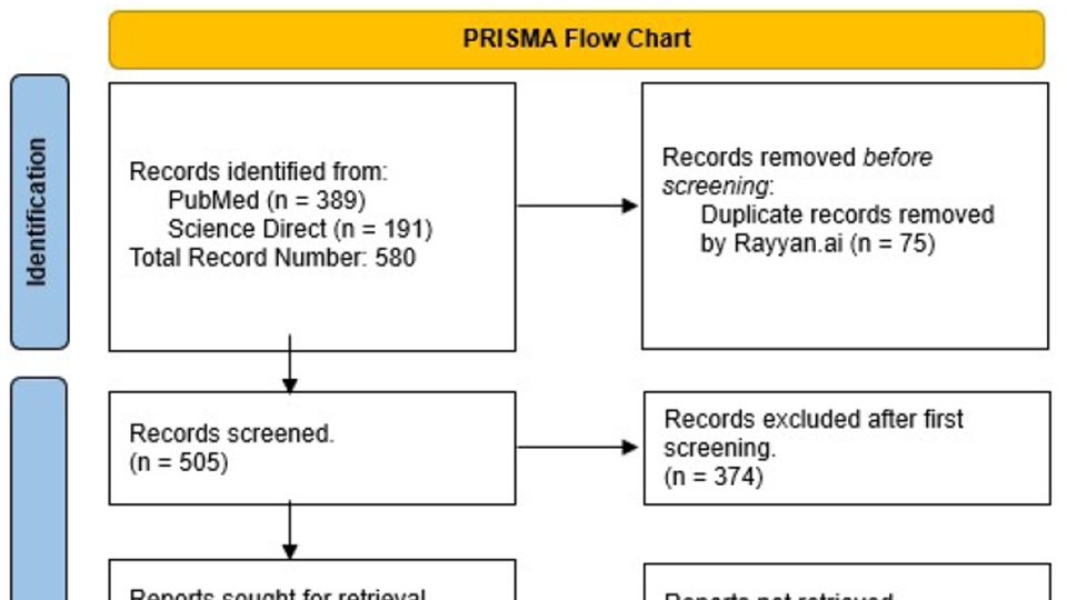
    Methods: Using the MeSH terms “Phage” “Therapy”, “Treatment” “Outcome”, a search was performed in ScienceDirect, Scopus and Pubmed databases, including narrative review, systematic reviews and clinical trials, dating from 2020 till June 2023. A total of 191 articles and 389 articles were retrieved respectively from each database, which after duplication removal, added up to 505. After the primary screen, 131 texts were collected. Following that, a secondary ended up with 56 articles included.

    Conclusion: With the constant increase in antibiotic resistance, there is a need for newer antimicrobial agents, which led to the revival of bacteriophages. Due to advances in molecular microbiology, phages became a possibility that has proven itself promising. More studies and funding are needed towards this field, that offers us a promising salvation from the antimicrobial resistance pandemic.


    Introduction:

         Bacteriophages, which are viruses that selectively infect bacterial cells, are ubiquitous in the environment and exhibit extensive morphological and genomic diversity 1. They possess basal plates with tail fibers that facilitate their initial attachment to host cells and introduce their genetic material via the sheath region. Bacteriophages follow a lytic or lysogenic life cycle within host bacterial cells, with the former resulting in the lysis of the host cell and the latter leading to the persistence of the bacteriophage in a latent state. Environmental cues, such as pH, nutrients, temperature, or exposure to antibiotics, can trigger prophages to enter a lytic cycle and the lysis of the host cell 1. Lytic bacteriophages are gaining popularity as a treatment for antibiotic-resistant bacterial infections, particularly the challenging 'ESKAPE' (Enterococcus faecium, Staphylococcus aureus, Klebsiella pneumoniae, Acinetobacter baumannii, Pseudomonas aeruginosa, and Enterobacter species) pathogens 1

         The “ESKAPE” pathogens are both Gram negative and positive species that are significant contributors to hospital-acquired infections, some of which are fatal 1. A recent study done by the US center for Diseases Control and Prevention, showed that we have roughly 51,000   health care associated infections reported to be caused by P.aeruginosa every year, 13% of these infections are MDR causing about 400 deaths per year. P.aeruginosa is found to have an increasing number of strains that are MDR without any effective antibiotic to stop this bacterium 2. The ESKAPE pathogen's capacity to evade the antibacterial effects of traditional antibiotic treatment renders the conventional therapy with antibiotics progressively less effective over time and raises the eminent need for alternative therapies, one of which is phage therapy 3.

         One application of phage therapy aims to target bacterial biofilms, which are a significant contributor to the antibiotic resistance of ESKAPE pathogens. In a study of 15 A.baumannii isolates, lytic phage reduced bacteria by 87%, indicating potential for breaking down biofilms on medical devices 4. However, phage treatment was not completely effective against all isolates, so combining multiple phages or phages with antibiotics may be necessary for optimal results 4. The "seesaw effect" is another mechanism of action observed in phage therapy. Here, induction of phage resistance leads to regained antibiotic susceptibility, highlighting an interesting evolutionary genetic trade-off. This suggests that using a combination of antibiotics and phages may be a beneficial approach when dealing with these superbugs 2.

         Phage therapy aims to provide personalized clinical treatment for patients. Successful trials include a 68-year-old patient with diabetes and necrotizing pancreatitis, and 4 severely ill COVID-19 patients with A.baumannii infection. Both trials showed positive results with complete resolution of A.baumannii infection demonstrating the significant potential of phage therapy for personalized or commercial use 5,6.  Similarly, case reports have shown the clearance of P.aeruginosa using phage therapy in several types of infections and has also been able to prove the safety of this therapy in a randomised, double blinded placebo-controlled clinical trial 7. Moreover, trials on bacteriophage therapy against acute and chronic S.aureus infections resulted in complete resolution of the diseases in 40% of patients after bacteriophage monotherapy, with better results (70%) after bacteriophage/antibiotic combination treatment 8.

         The main obstacle in evaluating the effectiveness of phage therapy in clinical settings is the limited number of trials due to the lack of regulations and safety guidelines, which prevent physicians from using them 9. Amongst the studies that were conducted, some notable limitations include a decreasing susceptibility to the phage over time, requiring multiple doses which was seen in the previously mentioned diabetic patient 5. Certain limitations are associated with the pathophysiology of specific pathogens, such as K.pneumoniae, which can reside inside cells within vacuoles, making it inaccessible to bacteriophages 10. Another is the development of anti-phage antibodies after oral phage therapy, which can reduce the efficacy of lytic phages. This was observed in patients with A.baumannii, K.pneumoniae, or P.aeruginosa infections after 2-3 weeks of phage therapy. Repeated phage treatment resulted in higher antibody levels, but its impact on phage therapy effectiveness requires further investigation 11.

         Bacteriophage therapy is superior to antibiotics usage as they generally possess self- replicating properties, selective targeting of a host, and lower side effects.1 Phage therapy can be utilized alone or in conjunction with antibiotics, and has demonstrated both efficacy and safety in clinical trials. However, certain limitations regarding clinical use and research remain, all of which will be discussed in detail in our review.

         This review is expected to provide a better understanding of phage therapy against ESKAPE organisms in terms of its effectiveness, indications of use and its anticipated side effects, by critically analyzing the existing literature and providing a comprehensive overview of this topic, in addition to identifying the current gaps in literature and providing new insights for future research for bacteriophages as a promising alternative to antibiotics in the treatment of bacterial infections.


    Methods

         This study reviewed the current knowledge regarding phage therapy in ESKAPE organisms, with particular emphasis on clinical indications, as well as therapeutic outcomes and side effects. In order not to exclude any important clinical article, a search strategy using the following principal MeSH terms was created: Phage Therapy, Treatment Outcome. The search was limited to ScienceDirect, Scopus and Pubmed databases, including narrative review, systematic reviews and clinical trials, dating from January 2020 till June 2023. A total of 191 articles and 389 articles were retrieved respectively from each database, which after duplication removal, added up to 505. Articles found in Scopus were duplicates from pubmed so they were automatically excluded. A primary screen was done through the article abstracts and title, and a total of 131 texts were collected. Following that, a secondary screening through the entire text was conducted, with a total of 56 articles were eventually included in the review.

    Study design:

    Narrative review

    Inclusion criteria:

    ●        Articles tackling MDR bacteriophage therapy for infections caused by ESKAPE microorganisms since the year 2020

    Exclusion criteria:

    ●        Articles in foreign language

    ●        Phage therapy in the context of oncologic & inflammatory non-infectious conditions

    ●        Studies dealing with non-human subjects

    ●        Studies done in laboratory settings (in-vitro)


    Results:

    Part I: General Overview:

         Multidrug resistant bacteria are becoming a major problem that the healthcare system is facing. Bacterial infections are slowly switching from curable to deadly diseases due to resistance 12. ESKAPE organisms are known to have different virulence factors and resistance abilities that makes them difficult to treat.  As these bacteria continue to develop resistance to almost all known antibiotics, shifting towards non-antibiotic interventions becomes a pressing need 13.

         Bacteriophages are types of viruses that are known to infect bacteria 14.  In the 1900’s, bacteriophage-based therapies were discovered and did show positive results for treating bacterial infection 14. D'Herelle's groundbreaking work in 1917, leading to the first application of phages as therapeutic agents in 1919 15. Back then, antibiotics were the most successful and safest agents available 14. The rise of antibiotics, providing a more convenient way to manage infectious diseases, overshadowed the phage therapy approach 15. Consequently, phage therapy was largely abandoned globally, except in a few countries like Georgia, Poland, and Russia, where it continued to be an approved treatment for specific bacterial infections 15. With the resurgence of interest in addressing bacterial infections, particularly due to multidrug resistance, there is a renewed focus on phage therapy as a potential solution 15.

         Being easily accessible with an acceptable safety profile, people misused antibiotics, leading to widespread antibiotic resistance. As this issue continued, pan-drug resistant organisms emerged and resistance became a global public health concern, persuading research to go to non-antibiotic agents to treat them 12.

         In fact, phages have been extremely effective against several types of infection, regardless of the pathogen itself. This is due to their ability to multiply specifically, which means that a lower therapeutic dose is needed to overcome the infection 16. In addition, resistance develops about ten times slower than antibiotic resistance 16. Our ability to genetically engineer viruses, provides us with a tool that can kill bacteria safely and effectively without many side effects that are seen with antibiotics 13.This is especially important in respiratory infections for example, that are a leading cause of morbidity and mortality 17.

         Nowadays, we can find numerous formulations for bacteriophage therapies, such as isolated purified phages, phage lysates, phage cocktails consisting of multiple distinct, and nanoparticles encapsulating phages for controlled release, along with phage-derived enzymes 15. The search for such alternative treatments gained traction from the 1980s to 1990s with antibiotic resistance, and phage isolation became a focal point for potential treatment options. However, challenges such as inadequate phage characterization, lack of controlled trials, and economic factors in assessment locations have hindered conclusive evidence of phage therapy's effectiveness 18.

         The cost of basic medical care is a significant concern in developing countries, exacerbated by the expense of acquiring effective antibiotics to combat antibacterial resistance 18. The potential of phage therapy in these areas is considerable, where infectious diseases cause millions of deaths annually. Phages could serve as a locally produced remedy, addressing antibiotic limitations and cost barriers 18.

         In recent years, the acceptance and application of phage therapy have increased in Western countries, paralleling Eastern countries where phages have been used clinically 18. Successful cases include patients infected with multidrug-resistant Acinetobacter baumannii and Pseudomonas aeruginosa. However, challenges have emerged, such as phage resistance and technical complexities in phage preparation 18.

    Part II: Advantages and Ease of Phage Utilization:

    Specificity

         There are several advantages of using therapeutic bacteriophages. For instance, phages exhibit a high degree of specificity, targeting only the harmful microorganisms while leaving beneficial microflora untouched 15. They are highly specific to their target bacteria and don't harm mammalian cells 3.  They usually target specific bacterial hosts based on the presence of complementary receptors on the bacterial surface 19. This specificity is advantageous as it allows for a narrow spectrum of action, reducing the impact on beneficial gut microbes and the development of secondary infections and antibiotic resistance 19. Studies have shown that phage therapy can be administered without disturbing the normal gut microbiome 19. This specificity contrasts with antibiotics that affect a broad range of bacteria, reducing the risk of complications like dysbacteriosis and secondary infections 15.

    Self-Replication

         Bacteriophages have a unique mechanism of action that allows them to lyse host targets and produce more phage copies, eliminating the need for repeated dosing or administration to maintain therapeutic concentration 19. This enhances their local antibacterial impact compared to antibiotics which require multiple dosing 15 19.

    Side Effect Profile

         Ad El Haddad et al. also demonstrated that administration of phages was generally well-tolerated, with no instances of adverse effects or toxicity reported 20. This was supported by many clinical trials 19. In addition, Phages have immunomodulatory properties, inhibiting inflammatory cytokine production and maintaining immune tolerance 19. Phages are generally safe when manufactured under Good Manufacturing Practice (GMP) guidelines, removing endotoxins and debris 21. Concerns arise from bacteriolysis events during phage administration, thus optimal dosing is crucial to prevent excessive cell wall fragmentation and cytokine increase 21. Al-Ishaq et al. reviewed studies demonstrated that various phage cocktails or individual phages were highly effective against pathogens and were considered safe, with no reported side effects 11.

    Resistance

         Resistance development, a major concern with antibiotics, is less problematic with phages due to their ability to coexist with bacteria and evolve to counter resistance 3. Even phage-resistant bacteria remain susceptible to other phages, and the process of introducing new phages is faster and more cost-effective compared to developing new antimicrobials according to regulations 15. Phages can reach infection sites effectively since they reproduce in the presence of their host bacteria and spread throughout the body when administered systemically 3. They can even penetrate areas like the blood-brain barrier, which may be inaccessible to drugs. Some phages can also disrupt biofilms. This growth behavior might allow for less frequent and lower treatment doses compared to antibiotics 3.

    Fighting Biofilms

         Some microbial agents produce biofilms, which makes them possess unique properties and resist conventional treatment methods 19. While antibiotics have limited effect against biofilm bacteria, bacteriophages have proven highly effective in eradicating them 19. Bacteriophages can infect and lyse biofilm-forming bacteria, degrade the EPS matrix, and penetrate through water channels within the biofilm structure 19. This is why phage therapy shows promise in treating persistent infections caused by biofilm formation on implanted medical devices 19.

        The use of bacteriophages for biofilm degradation is limited by their high host specificity. A phage can only eliminate its target bacterial species within a biofilm community, which is often composed of multiple bacterial species 19. While a cocktail of bacteriophages could be a solution, an alternative approach is the use of polyvalent phages with broad or multiple host ranges 19. Studies have shown the effectiveness of polyvalent phages in lysing mixed biofilms consisting of different bacterial species 19. This opens up opportunities for the expanded use of polyvalent phages in biofilm degradation, and there are numerous studies exploring their application in the literature 19.

    Cost Effectiveness

         Miedzybrodski et al. conducted a study involving six patients with antibiotic-resistant infections and compared the economic benefits of phage therapy with antibiotic therapy. The patients received oral phage preparation for 6.5 weeks, and the total cost of treatment was approximately 524 EUR 19. In comparison, the total cost of a ten-day course of vancomycin, linezolid, and teicoplanin ranged from 344 EUR to 2,620.85 EUR. Based on this analysis, the authors concluded that phage therapy was more cost-effective than antibiotic therapy for these infections 19.

    Chances for synergistic effect

         Applying a magnetic field can disrupt the chemical agents that hold microbial biofilms together 19. Studies have shown that antibiotics and magnetized bacteriophages can effectively penetrate and inhibit the growth of biofilms 19. This opens up new possibilities for using magnetic fields as an adjunct to antibiotic therapy and for applying bacteriophage therapy to eradicate drug-resistant bacterial infections caused by biofilm accumulation 19.

    Effective on Multi-Drug Resistant (MDR) bacteria

         Importantly, many of the bacterial strains used in the studies were resistant to commonly used antibiotics, making phage therapy a valuable alternative 11. In most cases, phages effectively reduced bacterial concentrations, improved outcomes, and provided protection against lethal infections or degraded biofilms 11.

         Antibiotic resistance can be decreased and improved antimicrobial effects are possible using phage-antibiotic therapy. According to studies, using phages in combination with antibiotics results in a lower antibiotic MIC, which helps remove biofilm 22. The best concentration of each antibiotic in a combination varies, though. Simultaneous administration produces synergism for aminoglycosides but not ciprofloxacin, demonstrating antibiotic class-specificity 22. Sequential delivery, for example with aminoglycosides, improves phage multiplication and bacterial death. The features of the host strain affect treatment effectiveness; antibiotic administration time and sequence affect synergistic effects. Due to phage specificity and antibiotic pharmacokinetics in physiological conditions, phage-antibiotic interaction results differ 22.

    Besides, bacteria may forgo virulence in favor of phage resistance, which could have positive effects including decreased antibiotic resistance and improved sensitivity to both phages and antibiotics. Recent research suggests that antibiotic susceptibility may increase in phage-resistant mutants, offering a technique to take advantage of bacterial weaknesses for effective phage therapy 23.

    Already Used and Proven Effective in Some Places

         In Egypt, Georgia, and the UK, phage therapy has shown promise in improving wound healing and recovery 24. Belgium also introduced a framework accepting phages as pharmaceutical ingredients 21. Phage potential to strengthen immune systems, lower bacterial loads, and treat pyoseptic issues in burn wounds has been demonstrated in clinical trials 34. Additionally, phages help wounds heal, fight off bacterial biofilms, and have transplant-safe immunosuppressive qualities 24. The benefits of phage therapy include its efficacy against drug-resistant germs, selectivity, and lack of hazardous side effects 24. Accurate agent identification, intracellular pathogen targeting, and routine phage efficacy monitoring present difficulties. Phage therapy offers a targeted and promising infection treatment strategy despite its difficulties 24.

    Support of Normal Flora Diversity

         The gut microbiome is significantly impacted by phage therapy. Phages support microbial diversity and guard gut mucosal surfaces 25. They can target problematic bacteria without disrupting the normal microbiome thanks to their restricted spectrum. According to research, phages may have an impact on diseases like diabetes and inflammatory bowel disease 25. However, it is unclear how they play a part in disease pathways. The effectiveness of fecal microbiota transplantation for illnesses like Clostridium difficile infection also appears to be influenced by phages 25. Phage therapy's effects on the gut flora and potential health advantages require further study 25.

    Effective Prototypes

         Modified S. carnosus phages, from an avirulent strain containing none of the virulence genes of S. aureus phages that could compromise the safe production of phages for therapeutic use, showed a wide host range, with rapid killing, high efficiency in reducing the formation of biofilms, and the prevention of appearance of resistant mutants, with only a small initiating dose required 26. Isolated lytic MRSA bacteriophages were shown to have wide host ranges, wide range of temperature, pH and still active at 2.5 kGy doses of gamma radiation, and forcing the MRSA strains to be sensitive after combination with vancomycin and erythromycin 27.


    Part III: Disadvantages and Ease of Phage Utilization

         The limitations of phage treatment can be broken down into three main categories: the effect of the phage's special qualities, the absence of suitable regulations, and standards, and the challenges with clinical applications 24. An ideal bacteriophage for therapeutic use should have good environmental adaptability and stability, be devoid of endotoxin genes in the genome, be easy to separate from other bacteriophages and to purify, and not harm the host 34.

    Lack of Adequate Research

         Despite the benefits of bacteriophage use, we still need to combat several difficulties to unlock their full potential. Clinical research and protocols for administration, dosage, frequency, and treatment duration are often lacking so far 3. The number of clinical trials assessing  the safety and indications of phage therapy in humans is very limited, and those that have occurred have involved small sample sizes and have relied on data generated by patients 9. The lack of appropriate legal and regulatory frameworks poses a significant challenge to integrating phage therapy into conventional medicine 3.

    Side Effects

         Antibiotics are known to have many side effects, but these effects are known, unlike those related to phage treatment 19. One very important hazard with phages is the risk of anaphylaxis due to the innate ability as viruses to induce an immune reaction 28. Viruses themselves can directly affect the immune system, triggering innate and adaptive responses 22. In addition, emergence of phage-neutralizing antibodies could hinder their effectiveness, as observed in some cases after parenteral administration 3. Other studies find the implications for such outcomes ambiguous 21.

         Besides, phage therapy can increase intestinal permeability and induce intestinal barrier dysfunction. Safety concerns in immunocompromised patients remain, but available literature suggests that phage therapy may be safe and effective for this population 19. Furthermore, phage-induced bacterial lysis can result in a rapid release of endotoxins, which may result in inflammatory reactions. It's crucial to keep track of the microorganisms present in phage preparations 22.

    Toxicity

         A lot of chemical excipients used with phage manufacturing can have their toxicities 29. Preparation often requires purification, often involving cesium chloride (CsCl), which is toxic 29. It is removed by anion exchange, which works well for smaller-scale purification22 .Although typically removed from clinical preparations, safer alternatives like polyethylene glycol, which are not free from toxicity, are used to mitigate risks 29.

         In addition to chemical toxicity, bacterial proteins and endotoxins present phage preparations may cause inflammation and hypotension. This is because when phages are prepared in the lab, bacteria are used to replicate them. For safety, it is essential to quantify toxin and bacterial particle presence 22.

    Bacterial Resistance

         Furthermore, bacteria can as well develop resistance to phages 30. Phages have the capacity to apply considerable evolutionary pressure on bacterial populations, prompting genetic alterations that lead to resistance 15. For instance, bacteria can employ diverse mechanisms to counteract phages, such as modifying their outer membranes, generating inhibitors that impede phage replication, or modifying the sites that phages target for infection 15. For example, P. aeruginosa can generate proteins that neutralize phage activity, enabling bacteria to flourish even in the presence of phages 15. Phage therapy typically involves using multiple phages to minimize resistance. Lysogenic phages, which incorporate their DNA into host bacteria, can potentially contribute to horizontal transfer of resistance genes19. However, lysogenic phages can also be modified to convey antibiotic-sensitive genes, offering a potential avenue for restoring antibiotic sensitivity in resistant bacteria 19. Nevertheless, phages can be used by bacteria as vectors to transmit their antibiotic and phage resistance genes via  genetic transduction, which can further spread resistance against them 28

    Host Specificity and Polymicrobial Infection

         The host specificity of phages can also be a limitation, particularly in polymicrobial infections and when broad-spectrum treatment is required 19. Large-scale production and distribution of phage-based therapeutics can be challenging due to regional specific epidemiology, as phages may be most effective when matched to the local microbial population 19. Understanding the regional specificity and targeting phages to prevalent antibiotic-resistant strains can improve the efficacy of phage therapy 19.

    Logistics

         Raising awareness of phage safety and efficacy is vital for clinical acceptance. Manufacturers must adhere to Good Manufacturing Practice guidelines to ensure phage stability from production to administration 20. Phages' personalized nature requires a distinct approach from traditional medicines, with considerations for quick updates in response to bacterial resistance 20. Phages also need for specialized expertise to administer them 15.

    Ideally, phage therapy offers a potential solution in the fight against multi-drug resistant bacteria in low-income countries, as effective and affordable alternatives to the very expensive and often unavailable antibiotics 19. However, limited financial resources, lack of political will, and regulatory limitations hinder the exploration of phage therapy in Africa 19.

    The adoption of phage therapy into conventional medicine faces regulatory hurdles due to the lack of a global framework. Its position as a personalized medicine approach challenges existing pharmaceutical regulatory policy 19. Issues related to intellectual property rights for phage therapy medicinal products (PTMPs) arise because natural bacteriophages cannot be patented, creating challenges for commercial production of phage cocktails 19. The need for market authorization for PTMPs is unclear, as some argue that they are formulated for specific patient use, while others require industrial-scale production 19. Additionally, the unique pharmacodynamic properties of phages as "active drugs" and their potential for self-replication in the body raise additional considerations 19. However, there is still much to learn from countries that have not abandoned phage therapy, particularly regarding research, development, policy, and legal considerations, if phage-based therapeutics are to be integrated into modern medicine 19.

    Examples from the Literature

         There are several case reports of unfavorable outcomes with phage treatment. One report discusses the development of reversible transaminitis in a patient with chronic methicillin-resistant S. aureus prosthetic joint infection following phage therapy was probably caused by underlying steatosis and cytokine response 31. Another describes a diabetic patient with necrotizing pancreatitis and Acinetobacter baumannii infection temporarily required more vasopressors following phage therapy, but things got better after phage therapy was resumed 22. During phage therapy, a child with P. aeruginosa bacteremia experienced anaphylaxis-related decompensation, possibly because of developing heart failure or endotoxin release 22. There is also a case of a patient with P.aeruginosa UTI who experienced fever and chills during phage therapy due to endotoxin release and the antibiotic regimen had to be changed 22.

         There are also many clinical trials involving phages. Phage therapy's safety was examined in trials for a variety of illnesses, including endocarditis, UTIs, venous leg ulcers, chronic otitis media, and more 22. A study in which healthy persons consumed E.coli phages revealed no changes in the amount of IL-4 in the blood, decreased fecal E.coli burdens, and no significant changes in the gut microbiota 22. In another study, healthy volunteers were given a broad-spectrum phage cocktail and experienced back discomfort, gastric acidity, allergic rhinitis, and low-grade fever 22. The PhagoBurn burn wound clinical trial examined phage therapy for burn wounds infected with P. aeruginosa. When a phage cocktail was given to the wound, the outcomes were disappointingly poor; this may have been because the phage dose was less than what was intended 23.

    Possible antagonistic effect with antibiotics

         Contrary to expectations, antibiotics like streptomycin caused bacterial resistance to phages 22. This is not only limited to streptomycin, as any antibiotic that interferes with protein translation and synthesis can stop the phage replication inside bacterial cells as well, limiting the awaited therapeutic effect 32.

    Part IV: Administration Route and Dosing

         The form and route of phage administration is a critical consideration for accurate dosage calculation 20. Some phages are used as ointments on wounds and burns. For that, gel applications have shown better outcomes in comparison to sprays 20. Oral delivery is challenging due to destruction in the stomach due to high acidity, but techniques like gastric fluid neutralization and capsule formulations can enhance absorption33. Intravenous administration is theoretically efficient but may trigger immune responses leading to fever or other constitutional symptoms 20.

         Current phage therapies don't have standardized formulations and are instead individualized. There are various methods, like as genetic engineering, phage bank expansion, and phage cocktail and antibiotic fusion 22. Although the development of topical and inhalable medications shows promise, there are still issues with dose, kinetics, and stability 22. The main challenge here is that phages are able to self-replicate, but if administered in low doses, bacteria can quickly resist them before they can overwhelm bacteria with their numbers. Besides, higher doses can have more side effects 33.

         Due to the synergistic effect of some antibiotics and phages, usage of such combos can be utilized to decrease the needed doses of both the phage and the antibiotic. However, it is worth mentioning that just as there exists no universal dose for all antibiotics, phages are very variant and no single dosing regimen fits them all 34.

    Part IV: The Emerging Problem of Multi-Drug Resistant Organisms (MDROs)

         As mentioned before, MDR emergence is a major challenge in infectious diseases. Pneumonias remain the most common cause to infection-relate mortality 35. The risks of death from pneumonia are augmented by antimicrobial resistance, especially in hospital-acquired and ventilator-associated settings 36.

         Solid organ transplant and hematopoietic stem cell transplant (HSCT) recipients are highly susceptible to infections and colonization by multidrug-resistant (MDR) pathogens. These MDR pathogens contribute to recurrent hospitalizations and infections, leading to a higher use of antibiotics 37. Bacterial prostatitis is also  becoming harder to treat due to resistance 38.

         The prevalence of MDR infections becomes major in the transplant population, with resistance can range from 14% in kidney transplant to 51% in lung transplant recipients 37. In HSCT patients, the prevalence of MDR pathogens can also reach up to 50%. These infections are associated with increased mortality and graft loss in transplant recipients 37.

         COPD and cystic fibrosis predispose to a infections that can be life-threatening and quickly gain resistance 39. These patients have high rates of colonization (up to 73%) with multidrug-resistant (MDR) pathogens, including Pseudomonas aeruginosa and Staphylococcus aureus 37. Besides, Burkholderia cepacia complex (BCC) species and Mycobacterium abscessus are associated with high rates of resistance 37. Lung transplantation is often contraindicated in patients with BCC or M. abscessus infections due to the high risk of recurrence and mortality 37. As these diseases become harder to combat, we move towards the “post-antibiotic” era” 12.

    A newly growing population, patients with ventricular-assist device (VAD) implantation have very frequent infections approaching 33% pr cases, often caused by Staphylococcus aureus and Pseudomonas aeruginosa37.

    Part V: Diseases with Phage Success:

         Phages offer a good antibiotic alternative, or antibiotic synergistic factor to help combar MDROs. For example, upon the oral administration of phage three times in individuals affected by bronchopneumonia empyema, 82% showed full recovery with a negative culture 8. The pressing need for rapid access to phage therapy in critically ill patients has prompted the initiation of compassionate use case studies in Western countries 21.

         It is known that phages can exist in their lytic or lysogenic phage and that they can both replicate in the targeted pathogen 40. Although both cycles can infect the bacterial host, only the lytic phage is used as “the phage therapy”, where the virus binds to the bacteria, injects its genetic material and replicate inside it until it bursts 40. Combining various phages with antibiotics to enhance their effectiveness against multidrug-resistant (MDR) bacteria and biofilms has gained significant attention. This strategy aims to augment antibiotic action by utilizing the phage ability to break down the extracellular polymeric substances (EPS) of biofilms and increase their permeability, enhancing antibiotic efficacy41.

         Tkhilaishvili et al. demonstrated that bacteriophages, administered alongside antibiotics, successfully inhibited MDR P. aeruginosa biofilm in a patient with diabetes, joint infection, and osteomyelitis 41. In another study, the combination of phage PEV20 and ciprofloxacin was employed to combat P. aeruginosa biofilm sourced from wound and cystic fibrosis patients. The study revealed that the joint application of antibiotics and phages exhibited superior biofilm eradication compared to the use of ciprofloxacin alone 42.

         Furthermore, phages are stable and can be stored at room temperature for months. They can survive stomach acidity, making them useful for treating intestinal colonization by bacteria like E. coli, Salmonella, Campylobacter, and Helicobacter 3.

         Many factors must be taken into account to optimize therapeutic outcomes. The patient's clinical status should guide the treatment duration, especially for those with multiple co-morbidities 20. For instance, patients with conditions such as diabetes mellitus, cystic fibrosis, obesity, smoking habits, or compromised immune systems might necessitate extended treatment periods 20. The severity and type of infection also play a role in determining treatment duration. Smokers, for instance, may require up to 18 weeks of phage therapy compared to non-smokers who might only need less than 4 weeks. However, immunocompromised patients might develop bacterial resistance after a few weeks of phage therapy, as observed in a lung transplant recipient with Pseudomonas aeruginosa pneumonia 20.

         Understanding Phage-Antibiotic Synergy (PAS) is pivotal for leveraging bacteriophage therapy in eradicating difficult to treat infections like osteoarticular infections 43. PAS has been demonstrated to curb the development of multi-drug resistant organisms by employing bactericidal mechanisms 43.

         There have been reports of successful outcomes in cystic fibrosis and lung transplant patients with MDR infections when treated with bacteriophage therapy in addition to systemic antibiotics 37. Case reports have also shown the safety and probable efficacy of intravenous bacteriophage therapy in pretransplant and posttransplant settings, with some patients also receiving nebulized phage 37.

         Some reports highlight successful outcomes in the treatment of extended-spectrum β-lactamase-positive Klebsiella pneumoniae infections 37. In one case, a kidney transplant recipient with recurrent urinary tract infection and epididymitis was treated with a combination of phages, administered orally and directly into the bladder via a urinary catheter for 12 weeks, alongside antibiotics. Another case involved a nontransplant recipient with a recurrent UTI caused by an extensively drug-resistant K. pneumoniae strain 37.

         Phage therapy also shows promise in treating infections in burn patients, by tackling problems brought on by bacteria that are resistant to antibiotics 24.

         Many other case reports on successful phage treatments exist. P. aeruginosa bacteremia was detected in a youngster who had Pseudomonas sepsis, where a phage cocktail was used when antibiotics proved ineffective to lessen the infection in the blood but not completely eradicate it 23. In another report, multiple phage cocktails were utilized in a case of necrotizing pancreatitis caused by an MDR A. baumannii-infected pseudocyst. A novel mixture proved successful in treating the infection over a 12-week period despite the emergence of resistance 23. P. aeruginosa was also targeted in an aortic graft: This infection proved resistant to conventional therapy. The local administration of ceftazidime along with phage OMKO1 successfully resolved the infection 23.

         Successful cases show how well phage treatment and antibiotics work together. Using a phage cocktail and trimethoprim-sulfamethoxazole, a patient with a UTI caused by the highly drug-resistant Klebsiella pneumoniae was able to get rid of the infection and avoid recurrence for six months 44. Similarly, a Georgia phage in combination with meropenem successfully treated an infection in a renal transplant patient, resulting in symptom relief and a year-long recovery.

         Depending on the kind, different phages and antibiotics interact differently. 44. S. aureus phages can also be used as a coating on prosthetic medical devices, in combination with antibiotics, as anti-biofilm formation strategies 45.

         Clinical trials have also been conducted on this topic. The Otitis Clinical Trial started in 2009 showed that phage treatment for persistent P. aeruginosa otitis dramatically lowered bacterial counts after treatment for 23 days. No significant adverse events were seen, and safety was established 23.

    In another trial in Bengladesh, phage mixtures were used to treat pathogenic E.coli in cases of diarrhea and failed to show benefot. The low-pH stomach environment may have an effect on viral viability, 23.

    Part VIL: Real Life Applications
         In a systemic review published in 2022, from 27 chosen studies that encompassed a patient cohort of 165 individuals, the analyzed data showed that 85% of cases exhibited decreased bacterial loads or complete eradication, coupled with improved clinical indicators, while 15% (24 cases) saw no positive impact from Phage Treatment (PT). A mere 21% (35 patients) underwent combined PT and antibiotic treatment, achieving a 100% success rate. Among the remaining 79% who exclusively received PT, the success rate reached 81%. Notably, within the 27 examined studies, 6 reported instances of bacterial resistance to phages 46. 
    In another review, 5 randomized clinical trials were included. One focused on chronic otitis where BT yielded significant positive clinical results 29.  The other trials suggested that the bacteriophage concentration might have been too low to trigger a significant therapeutic effect, raising the need for dose optimization 29. Additionally, two trials exploring topical bacteriophage use yielded discouraging results due to phages' susceptibility to environmental factors 29. Variations in phage variants, conditions, concentrations, and approaches in these trials make it challenging to draw definitive conclusions about bacteriophage efficacy 29.
         The effectiveness of phage therapy relies heavily on selecting precise bacteriophages for individual bacterial species and strains due to their host cell specificity. This necessity underscores the need for extensive phage libraries to tailor treatments accordingly 46.
    Phages carry diverse proteins crucial for their replication cycle, encompassing functions from host recognition to cell lysis 47. Among these, phage-encoded proteins like receptor binding proteins, Virion-associated peptidoglycan hydrolases (VAPGHs), endolysins, anti-CRISPR proteins, spanins and holins are being explored as potential tools to combat antibiotic-resistant bacteria 47. The effectiveness of phage-encoded proteins as therapeutics relies on their serum half-life, with longer half-lives generally considered preferable 47. Besides, safety and cost-effectiveness are important considerations throughout the production process, including host selection, endotoxin removal, and regulatory approval 47.
         The proper selection of phages for clinical application emerges as a pivotal factor in the success of phage therapy. This selection process is influenced by factors such as the type of infection, the timing of treatment, the patient's clinical status, and the attributes of the phages themselves 20. The reviewed studies underscore that appropriate phage selection is an essential consideration for successful clinical phage therapy. Effective phages should possess traits such as safety, strict lytic behavior, polyvalency, stability, ability to replicate within the host, and compatibility within phage cocktails to curb bacterial resistance 20. The absence of significant side effects in the reviewed studies can be attributed to the inherent composition of phages, primarily composed of DNA and proteins that do not provoke allergic or toxic reactions in humans, and their targeted specificity towards specific bacteria 20. Ensuring phage purity and appropriate dilution further enhances their safety profile by eliminating potential immune reactions and constitutional symptoms arising from bacterial lysates or toxins 20. Moreover, the risk of virulence or drug resistance genes within phage genomes is mitigated by employing naturally occurring, strictly lytic phages, as they lack integration with bacterial genomes and the dissemination of harmful genes 20. 
         Industrial-scale phage production requires standardized methods and regulatory frameworks lack global consensus 18. Raising awareness about phages' natural presence, safety, and potential efficacy is therefore crucial. Manufacturers must follow protocols, and regulations should ensure phage stability 18. 
    To address resistance, genetically engineered phages are being developed to enhance effectiveness and decrease immunogenicity 43. Notably, the US FDA has approved phage therapy under the "Emergency Investigational New Drug Scheme," with the European Medicines Agency considering genetically modified phages as advanced therapeutic medicinal products 43. Purification and dilution methods further enhanced phage safety by removing bacterial toxins and lysates 11. To ensure phage stability, it is crucial to use strictly lytic phages that do not carry resistance genes 11. Genome sequencing can help identify the presence or absence of resistance elements, guiding appropriate phage selection 11. Genetically engineered phages offer customization options, and they have demonstrated efficacy in reducing bacterial titers. In cases of phage resistance, the use of polyvalent phage cocktails in alternation can be considered 11.
         The administration of compassionate phage therapy typically involves a collaborative approach, bringing together experts from various fields, including infectious disease specialists, microbiologists, phage biologists, and pharmacologists 21. A standardized protocol for phage selection, production, purification, dosing, and treatment methods could immensely facilitate the broader implementation of compassionate phage therapy on a global scale. This would streamline the process and provide more uniform and effective treatments for critically-ill patients 21.
         Topical phage products have emerged as some of the first to reach the market, with Gladskin (lysin SA.100) by Micreos gaining approval from the European Medicines Agency 21. Gladskin targets S. aureus for treating skin conditions like eczema, rosacea, psoriasis, and acne 21. Micreos is also developing endolysin XZ.700 for atopic dermatitis. iNtRON Biotechnology completed a phase I clinical study for intravenous N-Rephasin SAL200, demonstrating its tolerability and safety 21. The endolysin exebacase CF-301 by ContraFect is being evaluated in COVID-19 patients with persistent MRSA bacteraemia, showcasing its effectiveness against a broad range of S. aureus isolates, including biofilms 21.
         Combination therapy, involving the use of both antibiotics and phage therapy, has shown synergistic effects in treating bacterial infections 11. Sublethal concentrations of antibiotics can enhance the production of lytic phages, leading to faster destruction of bacterial cells 11. In the reviewed studies, combination therapy was tested for S. aureus infections, with one study including P. aeruginosa. The results demonstrated mostly synergistic effects, such as improved survival, protection from lethal infection, biofilm degradation, and reduced bacterial load 11. However, factors such as administration methods, dosage, and choice of antibiotic can influence the practicality and effectiveness of the approach 11. 
         Currently, there are two main approaches to bacteriophage therapy in the United States. The first approach is personalized phage therapy, where a large phage library is developed and maintained 37. The patient's bacterial isolate is tested against various phages in the library to select the most effective ones 37. These phages are then used to create a personalized combination for the patient. Initially, finding matching phages took time as they were isolated from environmental and sewage samples 37. However, now there are commercial entities and academic laboratories with large phage banks, reducing the time to find active options 37.
    The second approach is the use of a fixed phage combination as an off-the-shelf product. This involves developing a product with a predetermined combination of phages that are active against a broad range of bacterial strains within a specific species 37. Genetically modified phages that can overcome bacterial resistance mechanisms can also be explored for this purpose 37. While not all patients can be treated with this approach, the goal is to be able to treat a majority of patients with susceptible bacterial infections 37.
         Unlike antibiotics, a single phage dosage may be able to completely eradicate the bacterial population 48. However, the method phages are given out directly affects how effective they are as a treatment. High quantities of phages must be delivered near to the target pathogen using the right dose and delivery techniques in order to obtain the best results 48. 
    Antibiotic and phage interactions can have positive or negative consequences. Rifampicin exhibits antagonistic behavior with some phages by reducing bacterial growth and phage replication through the host RNA polymerase 44. Rifampicin can work better when combined with phages that have their own RNA polymerase, such as the P. aeruginosa phage KZ 44. 
    Utilizing a variety of phages from nature is essential to preventing the emergence of phage resistance. Phage cocktails, which are combinations of several viruses, are used to boost therapeutic efficacy and immune system evasion 49. These combinations are effective because they dramatically lower the development of multi-resistance because even if bacterial cells develop resistance to one phage, they remain susceptible to the others in the mixture 49.
         Various formulations of phages have been reported that have different routes of administration to enhance bioavailability and target specificity, including topical applications, liposomal entrapments, immobilization of active phages on bandages, hydrogels, prosthetic devices and nanofibers. Encapsulating the phages in the liposome improves gastric resistance and stability, and protects it from antibodies 45. 
         One of the modes of administration is through nebulizers, which permits topical delivery of phages directly to the site of infection in patients with cystic fibrosis or pulmonary bacterial infections. In non-cystic fibrosis patients, inhaling small particles such as dry powders can incite irritation and bronchoconstriction, and thus extra caution is needed 45. 
    In one study, upon five nebulized administrations of pyophage cocktail, which included an anti-Staphylococcal phage, the patient suffering from cystic fibrosis became culture negative 50. Pyo bacteriophage preparation plays a role in wound healing in patients with diabetic foot ulcers colonized with MRSA and fully eliminate the infection, with the condition that it must be preceded with removal of necrotic tissue and antiseptic solution that doesn’t affect the phage’s titers, and continuous monitoring and the stopping of treatment only after the bacteria has immensely decreased 50. Topical preparation of staphylococcal phage Sb-1 once a week for seven weeks was shown to be more effective in toe ulcers treatment than antibiotics, but with a delay caused by osteomyelitis. Trials have also shown that S. aureus phages as a treatment for chronic venous leg ulcers are safe to use and with minimal side effects 50. 

    Part VII: ESKAPE Organisms and Phages

         In the treatment of infections caused by ESKAPE pathogens, phages have shown promise. Studies have demonstrated the effectiveness of phages against pathogens like Enterococcus faecalis and Staphylococcus aureus, where bacteriophage-based drugs have halted bacterial growth in both planktonic and biofilm stages 3.

         Combining lytic enzymes with cell-penetrating peptides, such as homing peptides, has been explored to target intracellular pathogens like E. coli, S. aureus, and L. monocytogenes 47. Data has demonstrated the safety profile of certain endolysins, suggesting their potential for therapeutic use. Moreover, endolysins like lysostaphin can be modified to create lysibodies, aiding in the opsonization of pathogens like S. aureus47.

         El Haddad et al. assessed the present utilization of phages as a therapeutic approach in Western countries by a systemic review. The majority of the reviewed research demonstrated that various phages and phage cocktails exhibited significant effectiveness against certain ESKAPE pathogens, while also maintaining a high level of safety 20. Notably, all patients enrolled in the review possessed bacterial strains resistant to conventional antibiotics. In approximately 87% of these cases, bacteriophage therapy exhibited positive outcomes, leading to reductions in bacterial concentrations, biofilm degradation, wound healing, and overall improvements in health 20.

         There is ongoing interest in using nebulized phage for outpatient management of P. aeruginosa infections in cystic fibrosis patients, particularly during cystic fibrosis flares 37. Efforts are also underway to develop a BCC phage library and a mycobacterial phage library targeting M. abscessus, M. tuberculosis, and M. avium species 37.

         Other antibiotic-resistant respiratory infection-causing microbes like Acinetobacter, Achromobacter, Staphylococcus, and Burkholderia species have also been successfully treated with phage therapy 50. Aerosolized phage therapy has been shown to be effective against the Burkholderia cepacia complex in mice 50.

         Careful phage selection is essential for phage mixtures to work well. Due to their wide host range and quick lytic cycle, the Myoviridae family is the main focus of most cocktails used to treat staphylococcal infections 49. Products like Pyophage, which contains a myovirus and a podovirus, are examples of exceptions. Although aggressive variants of these phages can be exploited, siphoviridae family phages are less frequently used due to their temperate lifestyle 49.

    Staphylococcus Aureus

         Another member of the ESKAPE family is Staphylococcus aureus. S. aureus is one of the main global contributors to both community and hospital-acquired illnesses. It is a highly adaptable pathogen that causes a wide range of illnesses, from minor skin and soft-tissue infections to fatal invasive infections 26. Methicillin resistant and vancomycin resistant S. aureus strains have emerged , which led to the emergence of bacteriophages infecting S.aureus as a potential treatment 49.  The prevalence of S. aureus (SA) phages in the environment is widespread 45. These include Siphovoridae, a temperate bacteriophage, but which may turn virulent, and Myoviridae and Podoviridae, which are virulent ones, have a wide host range, and are more preferred as they ensure the death of the infected bacteria 49.

         The efficacy of SA phages in the treatment of human infections was first reported after the regression of an infection within only 24-48 hours after the SA phages’ local injection in a surgically open wound 51.

         Trials on bacteriophage therapy against acute and chronic staphylococcal infections resulted in complete resolution of the diseases in 40% of patients after bacteriophage monotherapy, with better results (70%) after bacteriophage/antibiotic combination treatment, with the bacteriophage therapy being efficient in children, especially infants (aged 1–12 months) with staphylococcal infections 8

    Klebsiella Pneumoniae

        K. pneumoniae is a gram-negative bacterium that causes nosocomial antimicrobial-resistant opportunistic infections, including wound infections, pneumonia, meningitis, septicemia, and UTI 21. Regardless of antibiotic therapy, these infections can have a high mortality rate due to its incidence in neonates and immunocompromised patients. Over the last few decades, the rise in antibiotic resistance has resulted in the emergence of multidrug-resistant (MDR) strains, especially the Carbapenem-resistant K. Pneumoniae (CRKP), which pose a significant challenge in the treatment of infections. Facing antibiotic resistance, alternative methods of treatment are needed such as phage therapy, a promising tool yet in need of more research 21,52.

        There are few trials where Klebsiella bacteriophages were used in human subjects in EU and UK though nearly all the trials done were successful to treat MDR Klebsiella. This small number of trials is because the use of bacteriophage by physicians is hindered by the lack of regulations and rules regarding their safety 9.

        There are many bacteriophages targeting different components of the K. pneumoniae, most of them were tested in vitro or on animal models. For example, bacteriophage TSK1 produced a depolymerase enzyme and targeted the biofilm's capsular polysaccharides, resulting in a 99-100% reduction in the in-vitro developed K. pneumoniae biofilm 1. Bacteriophage cocktail, targeting many MDR bacteria, can also be effective after only 45 minutes of injections of isolates of 6 pathogens (including K. pneumoniae). Likewise, injection of the cocktail treatment involving bacteriophage ECP311, KPP235, and ELP140 displayed a 100 % survival rate when injected into an infected larva 1.

        K. pneumoniae bacteriophages have different host ranges, for instance, phage φBO1E recognizes and targets specifically K. pneumoniae of the pandemic clonal group 258 (CG258) clade II lineage, rendering it a narrow spectrum bacteriophage 9. On the other hand, phage KPO1K2, specific for K. pneumoniae B5055, is also able to infect multiple strains of K. pneumoniae, as well as some E. coli strains and, therefore, has a relatively broader host spectrum. Phages with a narrow host range are inappropriate for presumptive or prophylactic treatment 9. Successful phage therapy in combination with antibiotics is demonstrated by clinical examples. A phage cocktail and antibiotics were used to treat a patient with a UTI caused by Klebsiella pneumoniae and eradicate the infection for six months 44. In another instance, K. pneumoniae infection responded favorably to a Georgia phage and meropenem, resulting in relief and a year-long recovery.

    Acinetobacter baumannii

         A. baumannii causes nosocomial infections notably ventilator-associated pneumonia, meningitis, catheter-associated urinary tract infections and surgical site infections 53. It has become a significant concern in hospital settings due to its. In recent years, the bacteria has become more resistant to Colistin, a last resort antibiotic, making it more difficult to treat 53. Hospital-acquired infections caused by A. baumannii pose a significant risk to patients and can result in increased healthcare costs 53.

         One target of phage therapy is the breakdown of bacterial biofilm, in one study that evaluated lytic phage on 15 isolates of bacteria 4. The study's data analysis revealed that the isolated lytic phage decreased by 87%. This finding indicates that bacteriophages can be valuable in breaking down biofilms on the surface of medical devices 4. Nonetheless, the phage was not entirely successful in removing the biofilms of some isolates. Thus, to achieve optimal results and completely eradicate biofilms, a combination of multiple lytic phages or a combination of phages with antibiotics could prove beneficial 4.

         The goal of phage therapy is to ultimately be used for personalized clinical treatment of patients. Multiple trials have attempted to do so, where phage therapy successfully eliminated the Acinetobacter pneumonia with full recovery 5. In another trial, phage therapy was administered to four severely ill COVID-19 patients with A. baumannii pulmonary infections. Out of the four patients, three showed positive clinical results and were discharged home, however one died of respiratory failure a month later 54.

         Clinical administration of phage therapy in A. baumannii patients has been safe so far, with reported side effects including hypotension after phage administration, without the need for vasopressor administration 55. Some notable limitations of phage treatment include a decreasing susceptibility to the phage over time, requiring multiple doses 5. Another is the development of anti-phage antibodies, which could deactivate the lytic phages and impede their efficacy. This anti-phage humoral immunity was evident after 2-3 weeks of oral phage therapy for patients with A. baumannii, K. pneumoniae, or P. aeruginosa infections. Subsequent administration of phage treatment (3-5 courses) resulted in an increase in the levels of antibodies. Interestingly, there is no clear link between the strength of the immune response against the phage and the effectiveness of the phage therapy, hence this requires further investigation into its clinical significance 21

    Pseudomonas aeruginosa

         Pseudomonas aeruginosa is capable of evading the immune system by using its antiphagocytic polysaccharide capsule, biofilm formation and intracellular survival, causing fatal bacteremia and pneumonia 11. It is capable of causing extensive tissue damage by using its virulence factors acquired in hospitals where it is known to be the most common cause of ventilator acquired pneumonia 2 (VAP).

         A recent study done by the US center for Diseases Control and Prevention (CDC), showed that there are roughly 51,000 health care associated infections reported to be caused by P. aeruginosa every year, 13% of these infections are multidrug resistant causing about 400 deaths per year. P. aeruginosa is found to have an increasing number of strains that are multidrug resistant without any effective antibiotic to stop this bacterium 2.

         As mentioned previously, the key to phage therapy is the binding of the phage to its receptor and this phage-receptor relationship is widely affected by the environment of the phage itself. The presence of the phage receptors is influenced by their lipid surroundings that will affect their structure and function 7.  A slight change in the receptor can prevent phage binding, which means that it will not allow the phage to act as a bactericidal agent 7.

         Nevertheless, case reports have shown the clearance of P.aeruginosa using phage therapy in several types of infections and has also been able to prove the safety of this therapy in a randomized, double blinded placebo-controlled clinical trial 7.

         Various studies have demonstrated the promising potential of using bacteriophages against multidrug-resistant (MDR) Pseudomonas aeruginosa infections, particularly in cases of chronic otitis media, cystic fibrosis, and burn wounds 2. These studies have emphasized that phages need sufficient exposure time to penetrate biofilm exopolysaccharides and effectively clear P. aeruginosa. Overall, these pre-clinical studies highlight the potential of bacteriophages as a therapeutic approach for controlling MDR P. aeruginosa infections, particularly those associated with biofilm formation. A Pseudomonas aeruginosa infection in an aortic graft was successfully treated with phage OMKO1 and ceftazidime 44. Phage-antibiotic interactions can be antagonistic or synergistic, as in the case of the P. aeruginosa phage KZ with rifampicin 44.

         Due to its antibiotic resistance, Pseudomonas aeruginosa is a primary target for respiratory phage therapy 50. Phage treatment has been used to successfully treat respiratory P. aeruginosa infections, including those in CF patients, with minimal side effects 50.

    Enterobacter & Enterococcus

         The upsurge of antimicrobial resistance with sophisticated mechanisms has challenged the current fight against multi-drug resistant bacteria, in particular to VRE (Vancomycin-Resistant Enterococcus). PlyV12 lysin has shown strong lytic activity in vitro against multiple bacteria, including VRE 56. Other phages of the Siphoviridae & Myoviridae have been successfully used in vitro to eradicate Enterococcus faecalis 57. The advent of phage therapy comes with an interesting evolutionary genetic trade-off in E. faecalis, whereby induction of phage resistance comes at the cost of regaining antibiotic susceptibility, in what has been described the “seesaw effect”. This possibly promotes the idea of antibiotic-phage combinations when handling these superbugs 58. Alternatively, studies demonstrating phage-resistant Enterococcus faecalis have been handled by isolating yet another lytic phage against the bacteria, proposing cocktails of mixed phages57.

         Most of what is known regarding efficacy of bacteriophage therapy on Enterobacter spp., a facultative anaerobic rod-shaped bacilli responsible for bacteremia & sepsis in the context of nosocomial infection, has been limited to animal studies 59. There have been novel investigations of two bacteriophages ELP140 & GAP161 on survival rates of infected G. mellonella larvaes with the genus, both resulting in complete survival rates 59. As colistin is being increasingly used as a last resort for antimicrobial resistance, an alternative in phage therapy is being considered to colistin-resistant bacteria, which would otherwise pose a health concern 60. Isolation studies have succeeded in identifying lytic myPSH1140 phage against four colistin-resistant Enterobacter spp6. Multi-drug resistant strains of Enterobacter are quite common in E. cloacae & E. aerogenes, with phages belonging to the Podoviridae & Myoviridae respectively having been isolated 57.

    Discussion
         In the recent years, many articles have emerged in the literature discussing phages as potential non-antibiotic antimicrobials that can offer an alternative treatment option for infections, especially MDRO-induced disease 61. These viruses have been a source of attraction for many researchers and drug developers, due to their sturdiness, bacterial host specificity and ability to self-replicate 62. This offers several advantages, like a minimal side effect profile due to extreme specificity, an ease of storage and a possible 1 dose regimen 63.
         With the increase in MDROs and the paralleled increased mortality due to infectious diseases, phage studies have shifted from basic science and animal-based studies to human-based trials 63. This led to a great abundance of new information, especially about effectiveness, side effects, form of administration and dosing. For example, we have learned that now there are nebulized, IV, oral and topical phages 64. We have also learned that they minimally affect the gut microbiome and that they can be very effective in treating bacteria, sometimes outstanding antibiotics 65. This becomes especially evident in pan drug resistance and biofilms where antibiotics are least effective 66. 
         However, with more phage utilization, we discover new obstacles. Finding the right dose is very difficult in the absence of sufficient data. Besides, while specificity is a phage’s biggest advantage, it can be its enemy, where slight target modification can lead to resistance. Bacteria can also produce restriction enzymes to fight off their own “viral parasites” 62. While this can be combatted with the administration of polyvalent phages, their non-self-proteins are almost always immunogenic, which can trigger an immune reaction towards them, rendering them ineffective 67. Besides, bacterial cells are the breeding milieu for these viruses and thus phage therapy purification is key to avoid co-administration of bacterial remnants to avoid severe reactions such as anaphylaxis. This is less of a problem with topical or inhaled phages. Another obstacle is oral phages overcoming the stomach acidity barrier, which can be done by using liposomes and/or special capsules 68. 
         The promising thing is that now we have two potential antimicrobial options: phages and antibiotics. This does not mean that one is to replace the other, but rather as an added weapon to our war on infectious diseases. Therefore, we can have phage-antibiotic combos that would have synergistic effects, maximizing the effect of phages and antibiotics and minimizing their doses and side effects 69. This is especially true with antibiotics that target cell structures such as the cell wall or cell membrane. It is worth noting, however, that phages rely on cell machinery to make their own proteins, and by such antibiotics that target protein synthesis can have antagonistic effects to phages 69. 
         We now have an arsenal of experimental phages, but the data regarding them is still lacking. We need more large, randomized trials on phages to fully establish their dosing, side effect profile and efficacy so that we can move to large phase III trials. This way, we would be proactive in our battles against antimicrobial resistance, especially after finding new antibiotic classes or agents becomes more of a challenge 70. With our current technology we have made huge steps in genome sequencing and molecular microbiology that we are, more than ever, capable of achieving that by manipulating viral genomes in the way we find appropriate 71. This is why this era of antimicrobials is progressing to what some describe as the “post-antibiotic” or the “bacteriophage” era 72.

    Conclusion
         With the constant increase in antibiotic resistance, there is a need for new agents to fight off MDROs and increase cure rates and survivability with less side effects. This led to the revival of an idea that was long suppressed due to the ease of access to antibiotics: bacteriophages. However, antibiotics are gradually being overthrown by bacteria and now we have the technology to venture to molecular microbiology, making phages a possibility that has proven itself promising on multiple occasions. Therefore, more studies and funding are needed towards this field, that offers us a promising salvation from the antimicrobial resistance pandemic.

    Declarations

    Author contributions

    All authors contributed equally and validated the final version of record.

     Conflicts Of Interest

    The Author(s) declare(s) that there is no conflict of interest.

     Funding

    This research received no specific grant from any funding agency in the public, commercial, or not-for-profit sectors.

     Registration

    No registration applicable

     Data availability statement

    The data that support the findings of this study are available from the corresponding author upon reasonable request.

     Ethical approval

    The work presented in this article goes in accordance with the Declaration of Helsinki in 1964. 


    References

    1.      Patil A, Banerji R, Kanojiya P, Koratkar S, Saroj S. Bacteriophages for ESKAPE: role in pathogenicity and measures of control. Expert Rev Anti Infect Ther. 2021;19(7):845-865. https://doi.org/10.1080/14787210.2021.1858800.

    2.      Chegini Z, Khoshbayan A, Taati Moghadam M, Farahani I, Jazireian P, Shariati A. Bacteriophage therapy against Pseudomonas aeruginosa biofilms: a review. Ann Clin Microbiol Antimicrob. 2020;19(1):45. https://doi.org/10.1186/s12941-020-00389-5.

    3.      Aloke C, Achilonu I. Coping with the ESKAPE pathogens: Evolving strategies, challenges and future prospects. Microb Pathog. 2023;175:105963. https://doi.org/10.1016/j.micpath.2022.105963.

    4.      Ebrahimi S, Sisakhtpour B, Mirzaei A, Karbasizadeh V, Moghim S. Efficacy of isolated bacteriophage against biofilm embedded colistin-resistant Acinetobacter baumannii. Gene Reports. 2021;22:100984. https://doi.org/10.1016/j.genrep.2020.100984.

    5.      Schooley RT, Biswas B, Gill JJ, Hernandez-Morales A, Lancaster J, Lessor L, et al. Development and Use of Personalized Bacteriophage-Based Therapeutic Cocktails To Treat a Patient with a Disseminated Resistant Acinetobacter baumannii Infection. Antimicrob Agents Chemother. 2017 Sep 22;61(10):10.1128/aac.00954-17. https://doi.org/10.1128/aac.00954-17.

    6.      Wu N, Dai J, Guo M, Li J, Zhou X, Li F, et al. Pre-optimized phage therapy on secondary Acinetobacter baumannii infection in four critical COVID-19 patients. Emerg Microbes Infect. 2021;10(1):612-618. https://doi.org/10.1080/22221751.2021.1902754.

    7.      Lyon R, Jones RA, Shropshire H, Aberdeen I, Scanlan DJ, Millard A, et al. Membrane lipid renovation in Pseudomonas aeruginosa - implications for phage therapy? Environ Microbiol. 2022;24(10):4533-4546. https://doi.org/10.1111/1462-2920.16136.

    8.      Kwiatek M, Parasion S, Nakonieczna A. Therapeutic bacteriophages as a rescue treatment for drug-resistant infections - an in vivo studies overview. J Appl Microbiol. 2020;128(4):985-1002. https://doi.org/10.1111/jam.14535.

    9.      Herridge WP, Shibu P, O’Shea J, Brook TC, Hoyles L. Bacteriophages of Klebsiella spp., their diversity and potential therapeutic uses. J Med Microbiol. 2020;69(2):176-194. https://doi.org/10.1099/jmm.0.001141.

    10.  Cano V, March C, Insua JL, Aguiló N, Llobet E, Moranta D, et al. Klebsiella pneumoniae survives within macrophages by avoiding delivery to lysosomes. Cell Microbiol. 2015 Nov 1;17(11):1537–60. https://doi.org/10.1111/cmi.12466.

    11.  Al-Ishak R, Skariah S, Busselberg D. Bacteriophage Treatment: Critical Evaluation of Its Application on World Health Organization Priority Pathogens. Viruses. 2020;13(1):51. https://doi.org/10.3390/v13010051.

    12.  Royer S, Morais AP, da Fonseca Batistão DW. Phage therapy as strategy to face post-antibiotic era: a guide to beginners and experts. Arch Microbiol. 2021;203(4):1271-1279. https://doi.org/10.1007/s00203-020-02167-5.

    13.  Satta G, O’Callagharn C, Clokie MRJ, Di Luca M. Advancing bacteriophages as a treatment of antibiotic-resistant bacterial pulmonary infections. Curr Opin Pulm Med. 2022;28(3):225-231. https://doi.org/10.1097/MCP.0000000000000864.

    14.  Kuźmińska-Bajor M, Śliwka P, Ugorski M, Korzeniowski P, Skaradzińska A, Kuczkowski M, et al. Genomic and functional characterization of five novel Salmonella-targeting bacteriophages. Virol J. 2021 Sep 8;18(1):183. https://doi.org/10.1186/s12985-021-01655-4.

    15.  Baral B. Phages against killer superbugs: An enticing strategy against antibiotics-resistant pathogens. Front Pharmacol. 2023;14:1036051. https://doi.org/10.3389/fphar.2023.1036051.

    16.  Abedon S. Phage therapy pharmacology: calculating phage dosing. Adv Appl Microbiol. 2011;77:1-40. https://doi.org/10.1016/B978-0-12-387044-5.00001-7.

    17.  Wang R, Han JH, Lautenbach E, Tamma PD, Thom KA, Alby K, et al. Clinical prediction tool for extended-spectrum beta-lactamase-producing enterobacterales as the etiology of a bloodstream infection in solid organ transplant recipients. Transpl Infect Dis. 2021 Aug 1;23(4):e13599. https://doi.org/10.1111/tid.13599.

    18.  Kassa T. Bacteriophages Against Pathogenic Bacteria and Possibilities for Future Application in Africa. Infect Drug Resist. 2021;14:17-31. https://doi.org/10.2147/IDR.S284331.

    19.  Adesanya O, Oduselu T, Akin-Ajani O, Adewumi OM, Ademowo OG. An exegesis of bacteriophage therapy: An emerging player in the fight against anti-microbial resistance. AIMS Microbiol. 2020;6(3):204-230. https://doi.org/10.3934/microbiol.2020014.

    20.  El Haddad L, Harb CP, Gebara MA, Stibich MA, Chemaly RF. A Systematic and Critical Review of Bacteriophage Therapy Against Multidrug-resistant ESKAPE Organisms in Humans. Clin Infect Dis. 2019;69(1):167-178. https://doi.org/10.1093/cid/ciy947.

    21.  Chang RYK, Nang SC, Chan HK, Li J. Novel antimicrobial agents for combating antibiotic-resistant bacteria. Adv Drug Deliv Rev. 2022;187:114378. https://doi.org/10.1016/j.addr.2022.114378.

    22.  Liu D, Van Belleghem JD, de Vries CR, Burgener E, Chen Q, Manasherob R, et al. The Safety and Toxicity of Phage Therapy: A Review of Animal and Clinical Studies. Viruses. 2021;13(7):1268. https://doi.org/10.3390/v13071268.

    23.  Kortright KE, Chan BK, Koff JL, Turner PE. Phage Therapy: A Renewed Approach to Combat Antibiotic-Resistant Bacteria. Cell Host & Microbe. 2019;25(2):219-232. https://doi.org/10.1016/j.chom.2019.01.014.

    24.  Leontyev АE, Pavlenko IV, Kovalishena ОV, Saperkin NV, Tulupov АА, Beschastnov VV. Application of Phagotherapy in the Treatment of Burn Patients (Review). Sovremennye Tekhnologii V Meditsine. 2021;12(3):95-103. https://doi.org/10.17691/stm2020.12.3.12.

    25.  Guo Z, Lin H, Ji X, Yan G, Lei L, Han W, et al. Therapeutic applications of lytic phages in human medicine. Microb Pathog. 2020 May 1;142:104048. https://doi.org/10.1016/j.micpath.2020.104048.

    26.  Whittard E, Redfern J, Xia G, Millard A, Ragupathy R, Malic S, et al. Phenotypic and Genotypic Characterization of Novel Polyvalent Bacteriophages With Potent In Vitro Activity Against an International Collection of Genetically Diverse Staphylococcus aureus. Front Cell Infect Microbiol. 2021;11. https://doi.org/10.3389/fcimb.2021.698909.

    27.  Mahmoud ERA, Ahmed HAH, Abo-senna ASM, Riad OKM, Abo-Shadi MMAA–R. Isolation and characterization of six gamma-irradiated bacteriophages specific for MRSA and VRSA isolated from skin infections. J Radiat Res Appl Sci. 2021;14(1):34-43. https://doi.org/10.1080/16878507.2020.1795564.

    28.  Walsh L, Johnson CN, Hill C, Ross RP. Efficacy of Phage- and Bacteriocin-Based Therapies in Combatting Nosocomial MRSA Infections. Front Mol Biosci. 2021;8:654038. https://doi.org/10.3389/fmolb.2021.654038.

    29.  Au TY, Assavarittirong C. Combating antimicrobial resistance: an evidence-based overview of bacteriophage therapy. Postgrad Med J. 2023 Jul 1;99(1173):654–60. https://doi.org/10.1136/postgradmedj-2022-141546.

    30.  Flodman K, Tsai R, Xu MY, Corrêa IR, Copelas A, Lee YJ, et al. Type II Restriction of Bacteriophage DNA With 5hmdU-Derived Base Modifications. Front Microbiol. 2019;10:584. https://doi.org/10.3389/fmicb.2019.00584.

    31.  Doub JB, Ng VY, Johnson AJ, et al. Salvage Bacteriophage Therapy for a Chronic MRSA Prosthetic Joint Infection. Antibiotics (Basel). 2020;9(5):241. https://doi.org/10.3390/antibiotics9050241.

    32.  Pons BJ, Dimitriu T, Westra ER, van Houte S. Antibiotics that affect translation can antagonize phage infectivity by interfering with the deployment of counter-defenses. Proc Natl Acad Sci U S A. 2023;120(4):e2216084120. https://doi.org/10.1073/pnas.2216084120.

    33.  Khatami A, Foley DA, Warner MS, Barnes EH, Peleg AY, Li J, et al. Standardised treatment and monitoring protocol to assess safety and tolerability of bacteriophage therapy for adult and paediatric patients (STAMP study): protocol for an open-label, single-arm trial. BMJ Open. 2022 Dec 1;12(12):e065401. https://doi.org/10.1136/bmjopen-2022-065401.

    34.  Liu C, Hong Q, Chang RYK, Kwok PCL, Chan HK. Phage–Antibiotic Therapy as a Promising Strategy to Combat Multidrug-Resistant Infections and to Enhance Antimicrobial Efficiency. Antibiotics (Basel). 2022;11(5):570. https://doi.org/10.3390/antibiotics11050570.

    35.  Fernández L, Cima-Cabal MD, Duarte AC, Rodríguez A, García-Suárez MDM, García P. Gram-Positive Pneumonia: Possibilities Offered by Phage Therapy. Antibiotics (Basel, Switzerland). 2021;10(8):1000. https://doi.org/10.3390/antibiotics10081000.

    36.  Wang X, Xie Z, Zhao J, Zhu Z, Yang C, Liu Y. Prospects of Inhaled Phage Therapy for Combatting Pulmonary Infections. Front Cell Infect Microbiol. 2021;11:758392. https://doi.org/10.3389/fcimb.2021.758392.

    37.  Aslam S. Bacteriophage therapy as a treatment option for transplant infections. Curr Opin Infect Dis. 2020;33(4):298-303. https://doi.org/10.1097/QCO.0000000000000658.

    38.  Kraemer SD. Chronic Bacterial Prostatitis Treatment & Management [Internet]. Medscape; 2023. Available from: https://emedicine.medscape.com/article/458391-treatment


    39.  Ding F, Han L, Xue Y, Yang IT, Fan XF, Tang R, et al. Multidrug-resistant Pseudomonas aeruginosa is predisposed to lasR mutation through up-regulated activity of efflux pumps in non-cystic fibrosis bronchiectasis patients. Front Cell Infect Microbiol. 2022 Jul 27;12:934439. https://doi.org/10.3389/fcimb.2022.934439.

    40.  Xiong S, Liu X, Deng W, Zhou Z, Li Y, Tu Y, et al. Pharmacological Interventions for Bacterial Prostatitis. Front Pharmacol. 2020;Volume 11-2020. https://doi.org/10.3389/fphar.2020.00504.

    41.  Tkhilaishvili T, Winkler T, Müller M, Perka C, Trampuz A. Bacteriophages as Adjuvant to Antibiotics for the Treatment of Periprosthetic Joint Infection Caused by Multidrug-Resistant Pseudomonas aeruginosa. Antimicrob Agents Chemother. 2019;64(1):e00924-19. https://doi.org/10.1128/AAC.00924-19.

    42.  Glonti T, Chanishvili N, Taylor PW. Bacteriophage-derived enzyme that depolymerizes the alginic acid capsule associated with cystic fibrosis isolates of Pseudomonas aeruginosa. J Appl Microbiol. 2010;108(2):695-702. https://doi.org/10.1111/j.1365-2672.2009.04469.x.

    43.  Jeyaraman M, Jain VK, Iyengar KP. Bacteriophage therapy in infection after fracture fixation (IAFF) in orthopaedic surgery. J Clin Orthop Trauma. 2022;35:102067. https://doi.org/10.1016/j.jcot.2022.102067.

    44.  Li X, He Y, Wang Z, Wei J, Hu T, Si J, et al. A combination therapy of Phages and Antibiotics: Two is better than one. Int J Biol Sci. 2021;17(13):3573–82. https://doi.org/10.7150/ijbs.60551.

    45.  Khullar L, Harjai K, Chhibber S. Exploring the therapeutic potential of staphylococcal phage formulations: Current challenges and applications in phage therapy. J Appl Microbiol. 2022;132(5):3515-3532. https://doi.org/10.1111/jam.15462.

    46.  Aranaga C, Pantoja LD, Martínez EA, Falco A. Phage Therapy in the Era of Multidrug Resistance in Bacteria: A Systematic Review. Int J Mol Sci. 2022;23(9):4577. https://doi.org/10.3390/ijms23094577.

    47.  Aslam B, Arshad MI, Aslam MA, Muzammil S, Siddique AB, Yasmeen N, et al. Bacteriophage Proteome: Insights and Potentials of an Alternate to Antibiotics. Infect Dis Ther. 2021 Sep 1;10(3):1171–93. https://doi.org/10.1007/s40121-021-00446-2.

    48.  Kiani AK, Anpilogov K, Dhuli K, Paolacci S, Benedetti S, Manara E, et al. Naturally-occurring and cultured bacteriophages in human therapy. Eur Rev Med Pharmacol Sci. 2021;25(1):101–107. https://doi.org/10.26355/eurrev_202112_27339.

    49.  Jurado A, Fernández L, Rodríguez A, García P. Understanding the Mechanisms That Drive Phage Resistance in Staphylococci to Prevent Phage Therapy Failure. Viruses. 2022;14(5):1061. https://doi.org/10.3390/v14051061.

    50.  Jones JD, Varghese D, Pabary R, Langley RJ. The potential of bacteriophage therapy in the treatment of paediatric respiratory infections. Paediatr Respir Rev. 2022;44:70-77. https://doi.org/10.1016/j.prrv.2022.02.001.

    51.  Ji J, Liu Q, Wang R, Luo T, Guo X, Xu M, et al. Identification of a novel phage targeting methicillin-resistant Staphylococcus aureus In vitro and In vivo. Microb Pathog. 2020 Dec 1;149:104317. https://doi.org/10.1016/j.micpath.2020.104317.

    52.  Li F, Tian F, Nazir A, Sui S, Li M, Cheng D, et al. Isolation and genomic characterization of a novel Autographiviridae bacteriophage IME184 with lytic activity against Klebsiella pneumoniae. Virus Res. 2022;319:198873. https://doi.org/10.1016/j.virusres.2022.198873.

    53.  Weinberg SE, Villedieu A, Bagdasarian N, Karah N, Teare L, Elamin WF. Control and management of multidrug resistant Acinetobacter baumannii: A review of the evidence and proposal of novel approaches. Infect Prev Pract. 2020;2(3):100077. https://doi.org/10.1016/j.infpip.2020.100077.

    54.  Wu N, Zhu T. Potential of Therapeutic Bacteriophages in Nosocomial Infection Management. Front Microbiol. 2021;12:638094. https://doi.org/10.3389/fmicb.2021.638094.

    55.  Ibrahim S, Al-Saryi N, Al-Kadmy IMS, Aziz SN. Multidrug-resistant Acinetobacter baumannii as an emerging concern in hospitals. Mol Biol Rep. 2021;48(10):6987-6998. https://doi.org/10.1007/s11033-021-06690-6.

    56.  Haddad S, Jabbour JF, Hindy JR, Makki M, Sabbagh A, Nayfeh M, et al. Bacterial bloodstream infections and patterns of resistance in patients with haematological malignancies at a tertiary centre in Lebanon over 10 years. J Glob Antimicrob Resist. 2021 Dec 1;27:228–35. https://doi.org/10.1016/j.jgar.2021.09.008.

    57.  Kaur G, Agarwal R, Sharma RK. Bacteriophage Therapy for Critical and High-Priority Antibiotic-Resistant Bacteria and Phage Cocktail-Antibiotic Formulation Perspective. Food Environ Virol. 2021;13(4):433-446. https://doi.org/10.1007/s12560-021-09483-z.

    58.  Morrisette T, Kebriaei R, Lev K, Morales S, Rybak M. Bacteriophage Therapeutics: A Primer for Clinicians on Phage-Antibiotic Combinations. Pharmacotherapy. 2019 Dec 24;40(2):153–168. https://doi.org/10.1002/phar.2358.

    59.  Cieślik M, Bagińska N, Górski A, Jończyk-Matysiak E. Animal Models in the Evaluation of the Effectiveness of Phage Therapy for Infections Caused by Gram-Negative Bacteria from the ESKAPE Group and the Reliability of Its Use in Humans. Microorganisms. 2021;9(2):206. https://doi.org/10.3390/microorganisms9020206.

    60.  Borysowski J, Weber-Dabrowska B, Górski A. Bacteriophage endolysins as a novel class of antibacterial agents. Exp Biol Med (Maywood). 2006;231(4):366-377. https://doi.org/10.1177/153537020623100402.

    61.  Kasman LM, Porter LD. Bacteriophages. In: StatPearls [Internet]. Treasure Island (FL): StatPearls Publishing; 2022. Available from: https://www.ncbi.nlm.nih.gov/pubmed/29630237

    62.  Li XY, Lachnit T, Fraune S, Bosch TCG, Traulsen A, Sieber M. Temperate phages as self-replicating weapons in bacterial competition. J R Soc Interface. 2017;14(137):20170563. https://doi.org/10.1098/rsif.2017.0563.

    63.  Alvi IA, Asif M, ur Rehman S. A single dose of a virulent bacteriophage vB PaeP-SaPL, rescues bacteremic mice infected with multi drug resistant Pseudomonas aeruginosa. Virus Res. 2021;292:198250. https://doi.org/10.1016/j.virusres.2020.198250.

    64.  Durr HA, Leipzig ND. Advancements in bacteriophage therapies and delivery for bacterial infection. Mater Adv. 2023;4(5):1249–57. https://doi.org/10.1039/D2MA00980C.

    65.  Chung KM, Nang SC, Tang SS. The Safety of Bacteriophages in Treatment of Diseases Caused by Multidrug-Resistant Bacteria. Pharmaceuticals. 2023;16(10):1347. https://doi.org/10.3390/ph16101347.

    66.  Liu S, Lu H, Zhang S, Shi Y, Chen Q. Phages against Pathogenic Bacterial Biofilms and Biofilm-Based Infections: A Review. Pharmaceutics. 2022;14(2):427. https://doi.org/10.3390/pharmaceutics14020427.

    67.  Van Belleghem JD, Dąbrowska K, Vaneechoutte M, Barr JJ, Bollyky PL. Interactions between Bacteriophage, Bacteria, and the Mammalian Immune System. Viruses. 2018;11(1):10. https://doi.org/10.3390/v11010010.

    68.  Międzybrodzki R, Kłak M, Jończyk-Matysiak E, Bubak B, Wójcik A, Kaszowska M, et al. Means to Facilitate the Overcoming of Gastric Juice Barrier by a Therapeutic Staphylococcal Bacteriophage A5/80. Front Microbiol. 2017;8:467. https://doi.org/10.3389/fmicb.2017.00467.

    69.  Łusiak-Szelachowska M, Międzybrodzki R, Drulis-Kawa Z, Cater K, Knezevic P, Winogradow C, et al. Bacteriophages and antibiotic interactions in clinical practice: what we have learned so far. J Biomed Sci. 2022 Mar 30;29(1). https://doi.org/10.1186/s12929-022-00806-1.

    70.  WHO Antibacterial Pipeline Team. Lack of innovation set to undermine antibiotic performance and health gains [Internet]. World Health Organization; 2022. Available from: https://www.who.int/news/item/22-06-2022-22-06-2022-lack-of-innovation-set-to-undermine-antibiotic-performance-and-health-gains.

    71.  Habusha M, Tzipilevich E, Fiyaksel O, Ben-Yehuda S. A mutant bacteriophage evolved to infect resistant bacteria gained a broader host range. Mol Microbiol. 2019;111(6):1463–1475. https://doi.org/10.1111/mmi.14231.

    72.  Jin Y, Li W, Zhang H, Ba X, Li Z, Zhou J. The Post-Antibiotic Era: A New Dawn for Bacteriophages. Biology (Basel). 2023;12(5):681. https://doi.org/10.3390/biology12050681.


  • Efficiency and relevance of post-exposure prophylaxis prescription by emergency physicians: a monocentric retrospective study

    Abstract

    Background : Preventing infection with HIV remains a major public health challenge. Exposed persons are offered support and quick access to the post-exposure prophylaxis (PEP) by hospital emergency services. This study aimed to investigate the accuracy of the prescription of emergency PEP for patients with sexual fluid exposure in a French emergency department (ED) by comparing data in medical files and actual prescriptions.

    Methods : We retrospectively collected data for patients consulting for sexual exposure in a single Parisian ED from 2015 to 2016. For each patient, researchers independently checked whether the emergency physician prescribed PEP according to French guidelines. Our primary outcome was the appropriateness of the emergency PEP prescription after sexual fluid exposure. We calculated the Cohen weighted kappa coefficient with its 95% confidence interval (CI) for determining the agreement in indication for PEP.

    Results: We included 346 patients in the analysis. Half of the patients were men who had sex with men (n=178). The most frequent sexual exposure was anal insertive or receptive (n=177; 51%). PEP was prescribed in 94% of cases (n=328). In 33 cases (10%) the indication for PEP was not clear, but PEP was prescribed in 17 cases (52%). The Kappa value for determining the indication for PEP was 0.55 (95% confidence interval 0.36-0.74), indicating poor agreement for prescription. The agreement was lowest for men who had sex with men: 0.29 (0.05-0.53).

    Conclusions: Strategies are needed to improve the relevance of the prescription of PEP in French EDs to avoid the excess secondary effects and cost.

    Keywords: Sexual exposure; emergency medicine; post exposure prophylaxis


    Introduction

    Preventing infection with HIV type 1 (HIV-1) and 2 (HIV-2) remains a major public health challenge [1]. Emergency post-exposure prophylaxis (PEP) is an antiretroviral therapy for people exposed to risk of HIV transmission. PEP should be taken as soon as possible and at the latest, within 48 hrs of exposure [2]. Exposed persons are offered support and quick access to the PEP by hospital emergency services as exposure to HIV occupational and nonoccupational), is frequently managed in hospital emergency departments (EDs) [3, 4]. Guidelines for initiating PEP in EDs are regularly update [5]. The recommendations in France do not differ from those for US, United Kingdom or Italy in terms of risk stratification, the PEP indication primarily is relying on the description of the source partner [6-8]. Since 2011, the number of new cases of HIV diagnosed in France has been stable, about 6.000 per year [9]. Although PEP has real clinical impact, its use has some secondary effects (i.e., liver toxicity, hypersensitivity reactions) [10,11]. As well, in France, the cost of one PEP kit is 800 to 1,000 Euros and completely financed by public funds. Despite the important role of EDs in quelling the HIV epidemic, studies of emergencyphysician prescribing practices is limited [12, 13]. To our knowledge no study has investigated emergency-physician practices in prescribing PEP in France. We hypothesized that emergency physicians over-evaluate the risk of transmission of HIV after nonoccupational body fluid exposure and therefore overprescribe emergency PEP. Our aim was to describe emergency PEP prescription after body fluid exposure (mainly sexual) in a single emergency department.


    Methods

    Study design and setting

    We performed a retrospective study, from January 1, 2015 to December 31, 2016 in a single ED in Paris, France. The ED where the study was led is part of a teaching hospital and receives 90,000 yearly visits. In 2011, the incidence of HIV in Paris and its suburbs was 39/100.000, higher than that in France [14]. In France, patients who consult in an ED for non-occupational body fluid exposure are assessed by emergency physicians and are prescribed PEP according to the French guidelines [15]. When indicated, the emergency physician prescribes emergency PEP for 5 days, blood tests during the consultation are not recommended. Each patient will then be referred for specific infectious diseases consultation (Centre Diagnostic Anonyme et Gratuit) at the end of the 5-day course. There, an infectious-disease specialist evaluates the need to continue the treatment for 28 days and performs blood tests, including HIV-1 and -2, hepatitis B and C virus and syphilis.

    Selection of participants

    In the evaluated ED, each patient who consults has an electronic medical record. Before discharge, each physician will complete the file and select a discharge diagnosis from a list based on the International Classification of Diseases codes. All pathologies for blood or body fluid exposure are classified under the same code: Z20.9 “subjects exposed to communicable disease without precision”. For the study period, we collected data from charts with a discharge diagnosis of blood or body fluid exposure [16]. We included patients ≥ 18 years old, consulting in the ED for non-occupational HIV exposure (sexual exposure). Therefore, the non-inclusion criteria were age < 18 years, patient left without being seen, patient known to have HIV infection, consultation not related to HIV exposure, patient direct discharge to the infectious disease consultation, and occupational exposure.

    Outcomes measures

    Our primary outcome was the appropriateness of the emergency PEP prescription after sexual fluid exposure. For each patient, we evaluated the appropriateness of the prescription by comparing the emergency physician’s decision, or not, to prescribe PEP according to the French PEP national guidelines. When the medical file was incomplete to allow judgment, we decided a priori to consider that PEP was indicated. Secondary outcome was to assess factors associated with non-respect of post-exposure prophylaxis (PEP) recommendations.

    Patients and sexual fluid exposure characteristics

    Two researchers (XE and AC) independently extracted the following data from the ED electronic medical record using a standardized form: 1) patient demographics data (age and sex); 2) consultation details (day of consultation, period of consultation [night shift: from 22:00 to 8:00 and day shift from 8:00 to 22:00] and length of stay); 3) details related

    to the potential HIV exposure; and 4) If PEP was prescribed by the physician. For characterized sexual fluid exposure, researchers collected 1) the sexual practice (heterosexual, men who had sex with men [MSM], multiple partners); 2) risk of hemorrhage (anal, vaginal or buccal); and 3) if known, the HIV status of the patients’ partner.
    For each included patient, the researchers independently checked whether the emergency physician prescribed PEP according to French guidelines (PEP prescription was recommended, to be considered in view of file data, or not recommended) (Table 1). When assessments differed, the item was discussed until consensus was reached. When needed, a third reviewer assessed the report to achieve consensus. 

    Statistics

    Statistical analysis involved use of SAS 9.3 (SAS Inst. Inc., Cary, NC). Data are described with mean (SD) for quantitative variables and number (%) for qualitative variables. We calculated the Cohen weighted kappa coefficient with its 95% confidence interval (CI) for determining the agreement in indication for PEP. A kappa of 1, 0.90–0.99, 0.80–0.89, 0.70–0.79, 0.60–0.69, and <0.60 is considered perfect, excellent, very good, good, moderate, and poor, respectively [17]. Moreover, we performed a multivariate analysis of factors associated with non-respect of PEP recommendations, estimating odds ratios  and 95% CIs. The study was developed and results are reported according to the guidelines on the Strengthening the Reporting of Observational Studies in Epidemiology (STROBE) [18].


    Results

    Demographic data

    During the study 2-year period, we analyzed 534 consultations for exposure to communicable diseases. Two-thirds of the consultations were for sexual exposure (n=346) (Figure 1). Most patients were male (n=293; 85%). The mean age was 32 years (SD 10). Sexual orientation for patients did not differ by age (Table 2).
    For 51% (n=178) of cases, the sexual orientation of patients was MSM or bisexual; for 34% (n=117), heterosexual men; and for 15% (n=51), heterosexual women. Most patients consulted during the day rather than during the night shift. Consultations did not differ between the week and weekend. The mean length of ED stay was 126 min (SD 95).

    Description of the sexual exposure and source of HIV

    Almost all sexual exposures were with a single partner (n=338; 98%). The most frequent sexual exposures were vaginal insertive (n=103; 30%), receptive anal (n=95; 28%) and insertive anal (n=82; 24%) (Table 3). Half of the sexual exposures involved a torn condom (n=174; 50%). Half of the exposures were due to lack of a condom (n=165; 48%): 34 cases for heterosexual men, 17 for heterosexual women and 67 for MSM or bisexuals. The male condom was the only kind of mechanical protection reported. Seven patients (2%) reported an intact condom and did not have risky sexual exposure. Eight exposures (2%) were in a context of sexual assault. The positive HIV status was known for 10% (n=34) of source partners: 26 were MSM or bisexual, 5 heterosexual men and 2 heterosexual women. The viral load was reported detectable in 4 cases (12%) and unknown in 30 (88%). For 18% of cases (n=59), the source partner was in a high-risk group. The most frequent risk was multiple partners declared by the source partner. Most consulting patients could not say if the source partner had a risk factor (i.e., HIV positive, sharing a syringe, from a country with > 1% prevalence of HIV).  

    PEP prescription

    PEP was prescribed for 328 cases (94%). In 33 cases (10%), the indication for prescription was clearly not recommended, but PEP was prescribed for 17 cases (52%). The only factor associated with the nonadherence to the guidelines was the description of anal sex (insertive or receptive) during the sexual exposure (p<0.01) (Table 4).

    For 79 cases (23%), the prescription was clearly recommended, and in all cases emergency physicians followed the recommendations. In 233 cases (67%), emergency physicians prescribed the PEP, but the medical files did not allow for concluding whether PEP was indicated or not. The most frequent missing data were the country origin of the patient, the HIV status and the use of intravenous drugs. Emergency physicians prescribed PEP in almost all of these cases (n=231; 99%). The Kappa value for interobserver agreement for determining when PEP was indicated was 0.55 (95% CI 0.36-0.0.74), for poor agreement. The agreement was lowest for MSM patients: 0.29 (0.05-0.53). Table 5 presents the details of agreement.


    Discussion

    Our objective was to investigate the efficiency and relevance of the prescription of emergency PEP with sexual fluid exposure in a French ED. The PEP prescription remains a problem in EDs and in particular when the patient consulting is a MSM or bisexual. In fact, in these cases, in our cohort, the accuracy of the prescription was poor.

    Our study is the first to examine PEP prescription for emergency non-occupational blood exposure in France. A recent US study investigated care discrepancies between occupational exposure and non-occupational exposure in Eds [19]. The authors found that, emergency physicians failed to provide PEP in 27.5% of cases of high-risk non-occupational exposure and prescribed PEP correctly in 72.5% of cases (95% CI 66.8-77.5%). In 2008, Merchant et al., reported that PEP prescription after blood or body fluid exposure was low overall and varied by type of exposure [20]. Perhaps lack of knowledge of the indications for PEP by ED clinicians or lack of hospital protocols for PEP might explain the low prescription of PEP, especially after non-occupational exposure.

    Our findings are partially in conflict with these studies. In fact, like them, we found that French emergency physicians were not efficient in PEP prescription. However, French emergency physicians seem to over- than under-prescribe PEP. In an emergency situation, with an anxious patient who had a sexual exposure, refusing PEP prescription could be difficult even if the exposure was safe (i.e., vaginal insertive practice without a condom with a source partner with negative rapid-HIV serology findings).

    ED accessibility combined with the need to rapidly initiate PEP ensures that the ED is an important resource for patients after a sexual exposure. Information on physician decisions in the care of patients potentially exposed to HIV has been provided by physician surveys [21]. Among emergency physicians who care for patients after non-occupational exposure, only 15% recommended PEP after unsafe sexual practices and injection drug use.

    The management of sexual exposure in EDs could be improved in several ways. First, the development of expert consultations could help emergency physicians decide to prescribe or not PEP. Indeed, a free phone consultation with an HIV expert has been developed, the Clinical Consultation Center (CCC) [22]. This service provides clinicians of all experience levels with prompt expert responses to questions about HIV testing and prevention, HIV treatment, HIV–hepatitis co-infection, pre-exposure prophylaxis, perinatal HIV, substance use management, and PEP. If needed, expert consultation is available through the national PEP hotline, at 888-448-4911. Second, as always, the education of emergency physicians needs to be the base. The creation of an emergency specialty in 2017 will be a good opportunity to improve knowledge of the management of fluid exposure [23]. With substantial effort to develop new strategies to prevent HIV infection, emergency physicians must know the molecules, indications and implications. In fact, the prevention strategy could be crucially changed with the development of pre-exposure prophylaxis in high-risk groups [24]. Use of one pill daily can prevent up to 92% of new infections; most recent data show a number needed to treat of 13 to prevent one HIV infection [25, 26]. The US Food and Drug Administration has approved the prescription of preexposure prophylaxis by EDs. However, this treatment is associated with high rates of gastrointestinal and renal adverse events. So, it cannot be overprescribed by emergency physicians. Also, in contrast to other countries, the adherence and follow-up of patients with PEP in France has never been studied.


    Limitations

    There were limitations of this study. First limitation of our study is that it was retrospective. Thus, our results would require a prospective evaluation to confirm the results. Moreover, patient and/or physician interpretations of events leading to potential HIV transmission may be incompletely reported and conversations during consultation may have been inadequately documented. Some medical files did not have sufficient information to judge the appropriateness of care, which may have influenced results. The experience and pressure medical professionals face when patients present to the ED plays an important role in the decision to prescribe or not PEP to such patients. As our study was a monocentric evaluation, these findings may not be generalizable, even in a French setting since the reality in different hospitals is different and the available resources and manpower are different. Finally, a follow up on the outcomes and the results of the patients who received PEP would have been interesting, to see if the Emergency physician made the right judgment call at the time.


    Conclusions 

    This study showed that emergency physicians in France over-prescribe PEP, which exposes patients to risk of adverse events, and society to economic consequences. EDs must develop new strategies to respect guidelines on its prescription.


    Declarations

    Author contributions

    Conception and design: AC, XE and YY; Acquisition of data, AC and XE; Analysis: AC; Interpretation of data: AC, XE, PP, DP and YY; Drafting the article: AC, XE, PP, DP and YY; Revising it critically for important intellectual content: AC, XE, PP, DP and YY; Final approval of the version to be published: AC, XE, PP, DP and YY.

    Conflicts Of Interest: The Author(s) declare(s) that there is no conflict of interest.

    Funding: This research received no specific grant from any funding agency in the public, commercial, or not-for-profit sectors. 

    Registration: No registration applicable

    Data availability statement: The data that support the findings of this study are available from the corresponding author upon reasonable request.

    Ethical approval: Ethical approval for this study was not required.


    References

    1.     UNAIDS. The gap report [Internet]. 2014 [cited 2017 Jun 9]. Available from: http://www.unaids.org/en/resources/documents/2014/20140716_UNAIDS_gap_report.

    2.     Spira A, Marx P, Patterson B, Mahoney J, Koup R, Wolinsky S, et al. Cellular targets of infection and route of viral dissemination after an intravaginal inoculation of simian immunodeficiency virus into rhesus macaques. J Exp Med. 1996 Jan 1;183(1):215–25. https://doi.org/10.1084/jem.183.1.215.

    3.     Hsieh Y, Kelen G, Laeyendecker O, Kraus C, Quinn T, Rothman R. HIV Care Continuum for HIV-Infected Emergency Department Patients in an Inner-City Academic Emergency Department. Ann Emerg Med. 2015 Jul;66(1):69–78. https://doi.org/10.1016/j.annemergmed.2015.01.001.

    4.     Chen G, Jenkins E. Potential work-related bloodborne pathogen exposures by industry and occupation in the United States part I: an emergency department-based surveillance study. Am J Ind Med. 2007 Mar;50(3). https://doi.org/10.1002/ajim.20431.

    5.     Groupe d’experts pour la prise en charge du VIH sous la direction du Prof. Philippe Morlat, et sous l’égide du CNS et de l’ANRS. Prise en charge des accidents d’exposition sexuelle et au sang (AES) chez l’adulte et l’enfant [Internet]. CNS / ANRS; 2017 Sep [cited 2021 Jan 3]. Available from: https://cns.sante.fr/wp-content/uploads/2017/10/experts-vih_aes.pdf.

    6.     Kuhar D, Henderson D, Struble, KA, Heneine, W, Thomas, V, Cheever, LW, et al. Updated US Public Health Service guidelines for the management of occupational exposures to human immunodeficiency virus and recommendations for postexposure prophylaxis. Infect Control Hosp Epidemiol. 2013 Sep;34(9):875–92. https://doi.org/10.1086/672271. Erratum in: Infect Control Hosp Epidemiol. 2013 Nov;34(11):1238. Dosage error in article text.

    7.     Cresswell F, Waters L, Briggs E, Fox J, Harbottle J, Hawkins D, et al. UK guideline for the use of HIV Post-Exposure Prophylaxis Following Sexual Exposure, 2015. International Journal of STD & AIDS. 2016;27(9):713–38. https://doi.org/10.1177/0956462416641813.

    8.     Antinori A, Di Biagio A, Marcotullio S, Andreoni M, Chirianni A, d’Arminio Monforte A, et al. Italian guidelines for the use of antiretroviral agents and the diagnostic-clinical management of HIV-1 infected persons. Update 2016. New Microbiol. 2017 Apr;40(2):86–98.

    9.     Cazein F, Le Strat Y, Sarr A, Ramus C, Bouche N, Pillonel J, et al. Dépistage de l’infection par le VIH en France, 2003-2015. Bull Epidémiol Hebd. 2016;(41-42):745–8.

    10.  Pavel S, Burty C, Alcaraz I, de la Tribonnière X, Baclet V, Ajana F, et al. Severe liver toxicity in postexposure prophylaxis for HIV infection with a zidovudine, lamivudine and fosamprenavir/ritonavir regimen. AIDS. 2007 Jan 11;21(2):268–9. https://doi.org/10.1097/QAD.0b013e328011aa35.

    11.  Dellamonica P, Katlama C, Lévy-Bachelot L, Daures JP, Finkielsztejn L. Abacavir (Ziagen®) use between 2003 and 2008 in France according to the electronic medical record NADIS®. Med Mal Infect. 2013;43(11):467–74. https://doi.org/10.1016/j.medmal.2013.09.012.

    12.  Kelen GD, Hsieh YH, Rothman RE, Patel EU, Laeyendecker OB, Marzinke MA, et al. Improvements in the continuum of HIV care in an inner-city emergency department. AIDS. 2016;30(1):113–20. https://doi.org/10.1097/QAD.0000000000000896.

    13.  Stanley K, Lora M, Merjavy S, Chang J, Arora S, Menchine M, et al. HIV Prevention and Treatment: The Evolving Role of the Emergency Department. Ann Emerg Med. 2017 Oct 1;70(4):562-572.e3. https://doi.org/10.1016/j.annemergmed.2017.01.018.

    14.  Institut National de la Veille Sanitaire (INVS). Incidence de l’infection par le VIH [Internet]. 2013. Available from: https://www.santepubliquefrance.fr/maladies-et-traumatismes/infections-sexuellement-transmissibles/vih-sida/donnees/#tabs.

    15.  Ministère de la santé. Prise en charge des situations d’exposition au risque viral [Internet]. 2016 [cited 2017 Nov 1]. Available from: http://www.sante.gouv.fr/IMG/pdf/18_Prise_en_charge_des_situations_d_exposition_au_risque_viral.pdf.

    16.  Hivinfo [Internet]. 2021. Non-occupational Post-Exposure prophylaxis (nPEP). Available from: https://hivinfo.nih.gov/understanding-hiv/fact-sheets/post-exposure-prophylaxis-pep.

    17.  Landis JR, Koch GG. An application of hierarchical kappa-type statistics in the assessment of majority agreement among multiple observers. Biometrics. 1977 Jun;33(2):363–74.

    18.  Von Elm E, Altman DG, Egger M, Pocock S, Gøtzsche P, Jan P, et al. The Strengthening the Reporting of Observational Studies in Epidemiology (STROBE) Statement: Guidelines for Reporting Observational Studies. Ann Intern Med. 2007;147(8):573–7. https://doi.org/10.7326/0003-4819-147-8-200710160-00010.

    19.  O’Donnell S, Bhate TD, Grafstein E, Lau W, Stenstrom R, Scheuermeyer FX. Missed Opportunities for HIV Prophylaxis Among Emergency Department Patients With Occupational and Nonoccupational Body Fluid Exposures. Ann Emerg Med. 2016 Sep 1;68(3):315-323.e1. https://doi.org/10.1016/j.annemergmed.2016.03.027.

    20.  Merchant RC, Becker BM, Mayer KH, Fuerch J, Schreck B. Emergency Department Blood or Body Fluid Exposure Evaluations and HIV Postexposure Prophylaxis Usage. Acad Emerg Med. 2003;10(12):1345–53. https://doi.org/10.1111/j.1553-2712.2003.tb00009.x.

    21.  McCausland JB, Linden JA, Degutis LC, Ramanujam P, Sullivan LM, D’Onofrio G. Nonoccupational postexposure HIV prevention: Emergency physicians’ current practices, attitudes, and beliefs. Ann Emerg Med. 2003;42(5):651–6. https://doi.org/10.1016/S019606440300338X.

    22.  Clinician Consultation Center [Internet]. 2016 [cited 2017 Sep 6]. PEP Guidelines. Available from: https://nccc.ucsf.edu/clinical-resources/pep-resources/pep-guidelines/

    23.  Riou B. 2017 : l’an 1 du diplôme d’études spécialisées de médecine d’urgence. Annales françaises de médecine d’urgence. 2017 Feb 1;7(1):1–4. https://doi.org/10.1007/s13341-017-0710-z.

    24.  Molina JM, Capitant C, Spire B, Pialoux G, Cotte L, Charreau I, et al. On-Demand Preexposure Prophylaxis in Men at High Risk for HIV-1 Infection. N Engl J Med. 2015;373(23):2237–46. https://doi.org/10.1056/NEJMoa1506273.

    25.  Grant RM, Lama JR, Anderson PL, McMahan V, Liu AY, Vargas L, et al. Preexposure Chemoprophylaxis for HIV Prevention in Men Who Have Sex with Men. N Engl J Med. 2010;363(27):2587–99. https://doi.org/10.1056/NEJMoa1011205.

    26.  McCormack S, Dunn DT, Desai M, Dolling DI, Gafos M, Gilson R, et al. Pre-exposure prophylaxis to prevent the acquisition of HIV-1 infection (PROUD): effectiveness results from the pilot phase of a pragmatic open-label randomised trial. Lancet. 2016 Jan 2;387(10013):53–60. https://doi.org/10.1016/S0140-6736(15)00056-2.

Latest articles

Loading articles...

Indexing Information

Loading metrics...

PoEM News

Loading news...

Partnership

Loading partnership...

Journal Metrics

Loading metrics...

Meet the Board

Support for Authors

1

Getting Started

Register with journal and receive Author role. Access dashboard to begin new submission.
?
2

Complete Submission

Select language, title, section • Enter abstract, keywords, references • Upload files • Add co-authors
?
3

Review & Submit

Add optional comments for editors and suggest reviewers. Review all information and confirm submission.
?

Submitted

Your submission is complete and under review

Published

Article successfully published
8

Copyediting

Review copyedited files and publication-ready versions. Provide feedback or approval through discussions.
?
7

Acceptance

Accepted manuscripts proceed to copyediting and production. Receive confirmation notifications.
?
6

Upload Revised

Upload revised files through Review tab with response to reviewers (if revisions required).
?
5

Peer Review

Receive editor's decision via email and dashboard.
?
4

Track Submission

Monitor submission status via Author Dashboard.
?

Support for Reviewers

1

Receive Invitation

Editors send invitation via email with manuscript details. Access via link or journal login.
?
2

Request Details

Click to access assignment details including article title, abstract, review files, and schedule.
?
3

Accept / Decline

Review submission details and privacy statement. Decide whether to accept or decline the assignment.
?
4

Read Guidelines

Review journal's specific reviewer guidelines. Guidelines remain accessible throughout the review process.
?
5

Review Manuscript

Download review files and conduct thorough review. Enter comments for editor and author.
?

Complete

Review successfully submitted
9

Submit Review

When satisfied with review and recommendation click Submit. Confirm submission to complete the process.
?
8

Communicate

Use Review Discussion panel to ask questions. Click Add discussion to start conversation.
?
7

Recommendation

Select recommendation from dropdown menu based on review: Accept, Revisions, Resubmit, or Decline.
?
6

Upload Files

Optionally upload marked-up copy of review file. Remember to anonymize if required by type.
?
PoEM Footer